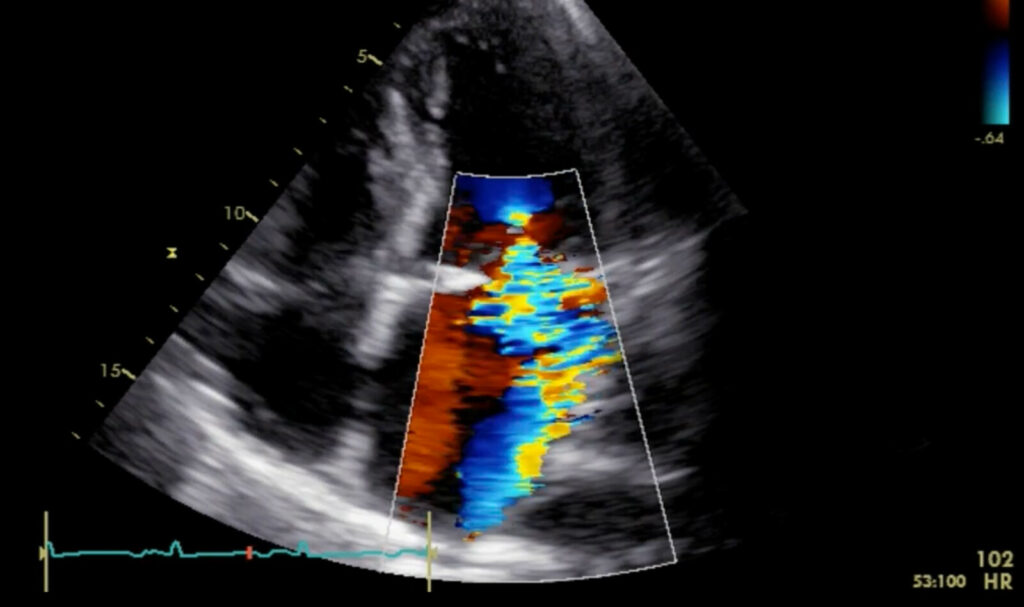
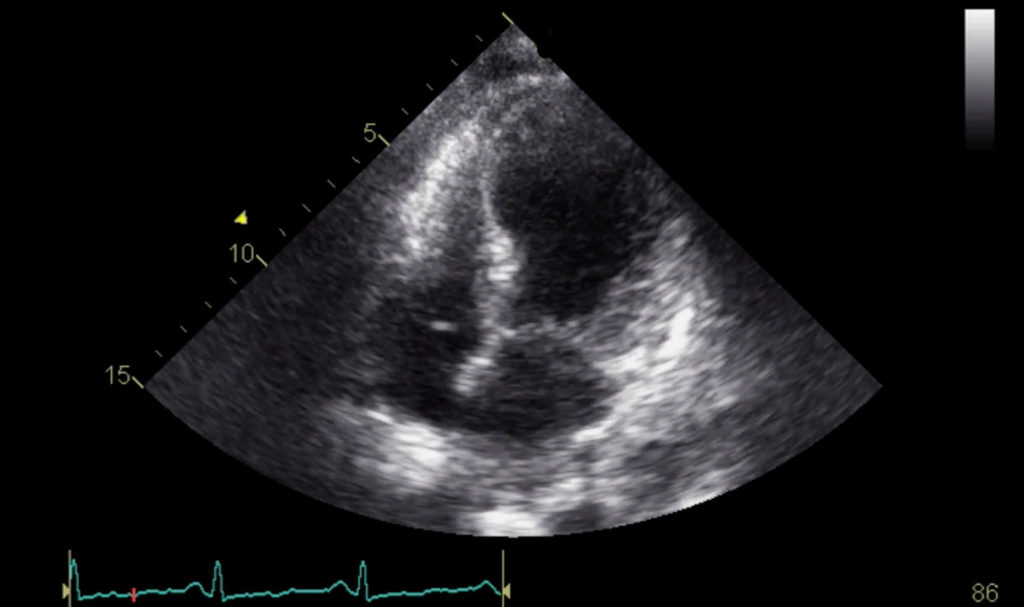
CLINICAL INVESTIGATIONS • Passive Leg Raise Stress Echocardiography in Severe Paradoxical Low-Flow, Low-Gradient Aortic Stenosis
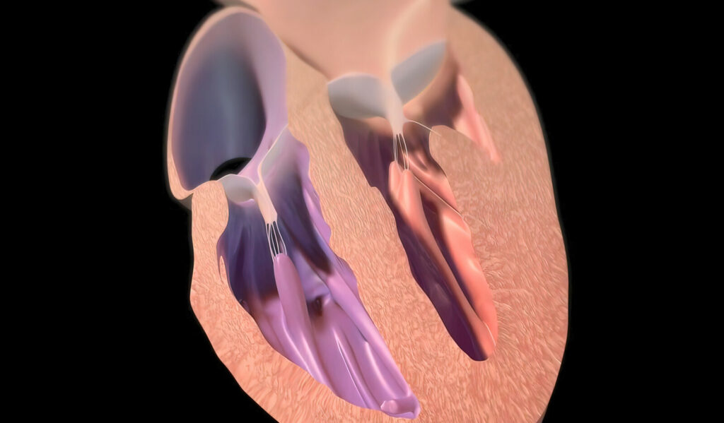
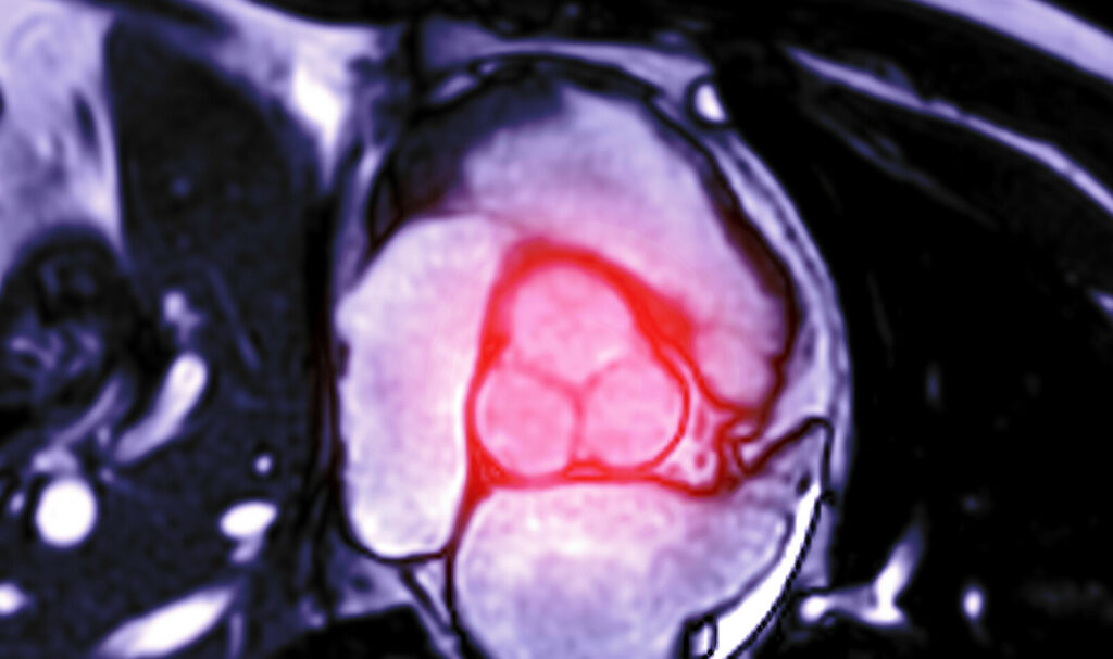
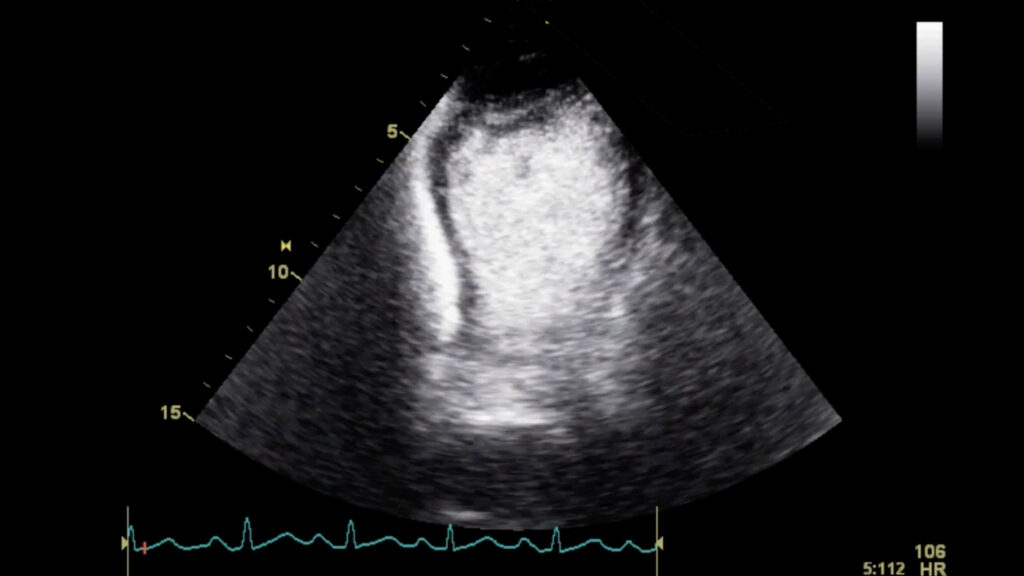
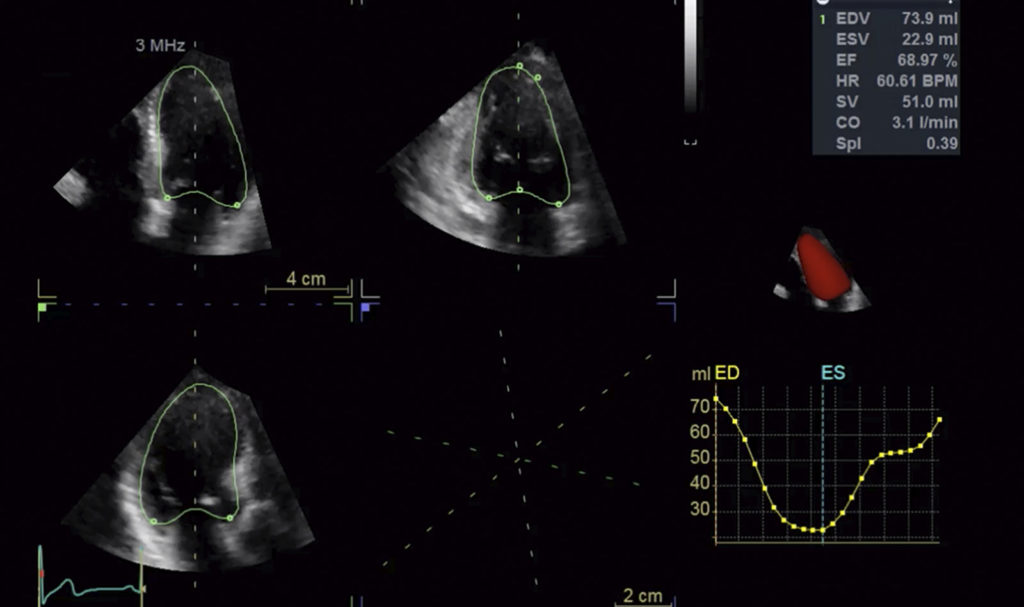
Management of aortic stenosis (AS) – the most common valvular heart disease in Europe and North America – is done by catheter intervention or surgery; in severe symptomatic AS, valve replacement is the treatment of choice. Diagnosis of severe AS, which is defined as an aortic valve area (AVA) ≤ 1.0 cm2, is done with echocardiography. The left ventricle (LV) ability to produce transvalvular flow that is sufficient in generating high gradients varies…
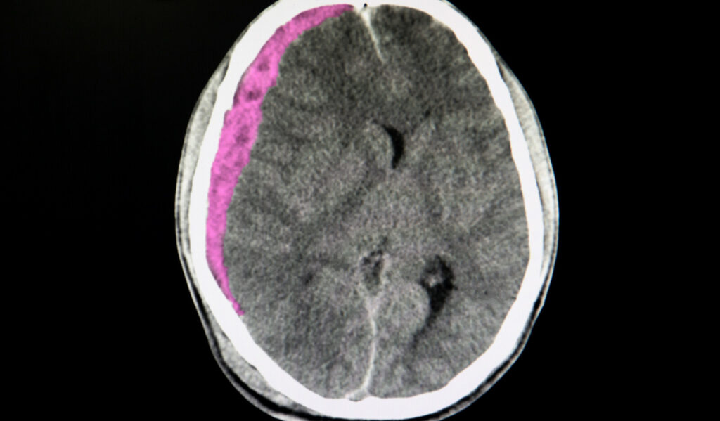
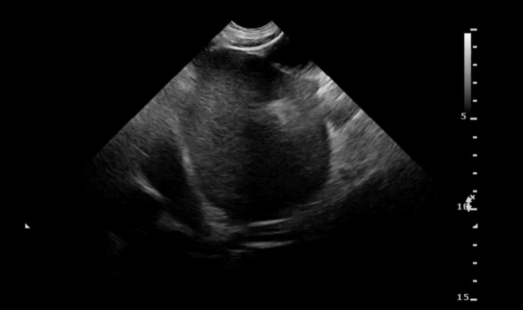
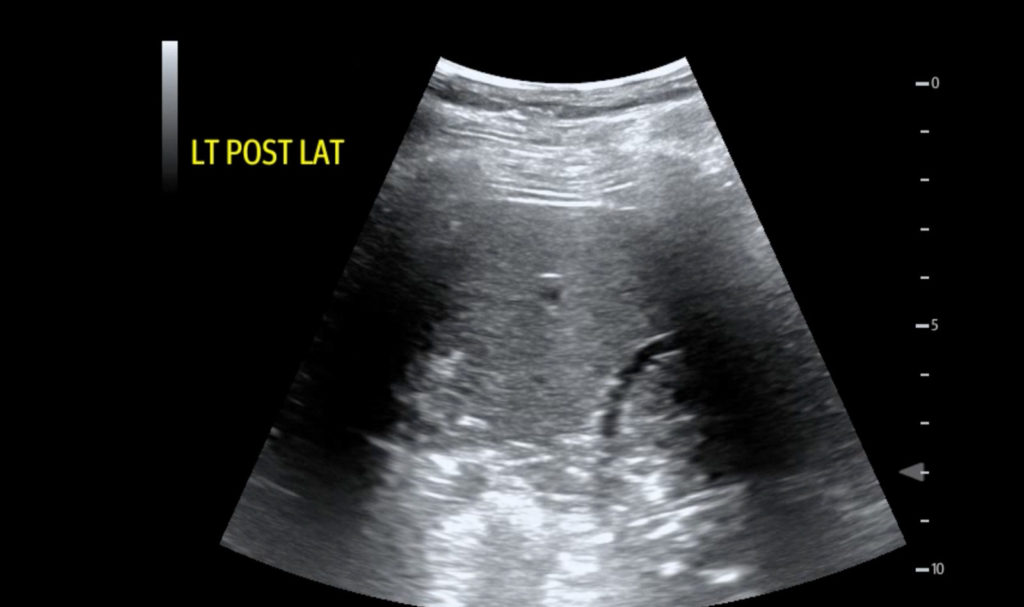
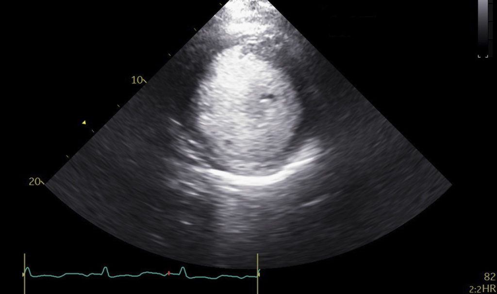
ORIGINAL ARTICLE • Exploratory Study to Assess Feasibility of Intracerebral Hemorrhage Detection by Point of Care Cranial Ultrasound.
Use of ultrasound for detection of intracerebral hemorrhage (ICH) using diagnostic high end transcranial color-coded duplex machines has been only limited studied. There is no published literature on imaging techniques and parenchymal topography for cranial B mode imaging. Studies assessing feasibility and accuracy of cranial POCUS to diagnose intracerebral pathology such as ICH are sparse.
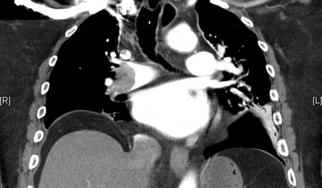
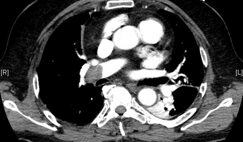
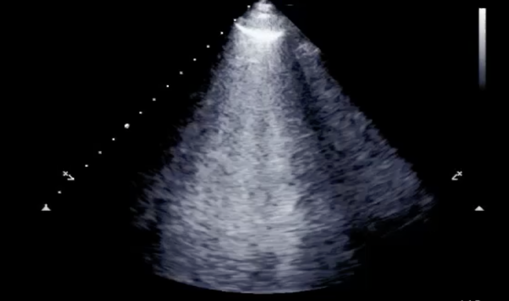
ORIGINAL ARTICLE • Retrospective Analysis of the Diagnostic Accuracy of Lung Ultrasound for Pulmonary Embolism in Patients with and without Pleuritic Chest Pain
Pleuritic chest pain – sharp chest pain exacerbated by breathing or coughing – is a common presenting symptom in the emergency department (ED) and requires a careful differential diagnosis between benign conditions such as musculoskeletal pain, and more serious diseases like pulmonary embolism (PE), pneumothorax, pneumonia with pleuritis and cancer. Among these conditions, PE is a major cause of morbidity, mortality, and hospitalization.
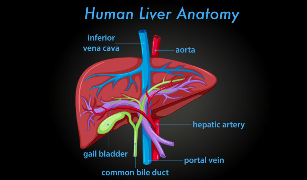
RESEARCH • Doppler Study of Portal Vein and Renal Venous Velocity Predict the Appropriate Fluid Response to Diuretic in ICU: A Prospective Observational Echocardiographic Evaluation.
There has been a growing interest in the diagnosis and treatment of fluid overload and venous congestion in recent years. Fluid overload implies peripheral edema but also could result in pulmonary edema and venous congestion, which can alter tissue perfusion. Several studies have shown an association between fluid overload and morbidity and mortality in intensive care unit (ICU).
eMedical Academy Releases Article Helping to Answer CCEeXAM Questions by Sharing What Past Test Takers Have to Say About the Exam
eMedical Academy, an online ultrasound school for Point-of-Care ultrasound continuing education, recently published a new piece on their website addressing CCEeXAM questions ultrasound professionals might have, by revealing feedback from students who have previously taken the test…
THE HEMODYNAMIC CORNER • Aortic Stenosis Severity: Rhythm Makes a Difference
The assessment of aortic valve stenosis (AS) severity based on Doppler velocity and pressure gradient is flow dependent. Both velocity and pressure gradient increase with elevated transaortic flows and decrease with reduction in flow rates for a given aortic valve orifice area. In this case report, the authors describe a challenging case of assessing AS severity in the setting of bradycardia from complete heart block (CHB), normal left ventricle (LV) function, permanent atrial fibrillation (AF), and moderate AS at baseline.
REVIEW • Cardiac Imaging for Diagnosis and Management of Infective Endocarditis
In-hospital mortality of infective endocarditis (IE) remains approximately 20%, and 1-year mortality approaches 40%. Decision-making in patients with IE is often complex, requiring collaboration among cardiologists, cardiac surgeons, and infectious disease specialists, among others. Although echocardiography has long been the principal imaging modality in this disorder, others including positron emission tomography (PET) and cardiac computed tomography (CT), and to a lesser extent intracardiac echocardiography (ICE), play an increasing role.
CCEeXAM Questions? Here’s What Past Students are Saying…
Have CCEeXAM questions? Let’s look at what past test takers have said about the CCEeXAM and how you can prepare for it and be confident….
eMedical Academy Publishes New Article Discussing Importance of CCEeXAM Prep and Why It Matters for Echocardiographers
eMedical Academy recently partnered with Inteleos to be one of their POCUS Education Provider Network. This network includes independent POCUS Education Providers who have been vetted by the Inteleos POCUS Certification Academy to provide quality education and training experience to new and advanced POCUS users…
CASE REPORT • Notching into a Diagnosis—Incorporating Doppler Interrogation into Point-of-Care Ultrasonography to Diagnose a Submassive Pulmonary Embolism.
Often, patients with pulmonary embolism (PE) present with nonspecific symptoms. This, in turn, pose a diagnostic dilemma. The authors present in this case report a case of submassive PE diagnosed in a patient utilizing point-of-care ultrasonography (POCUS) in the emergency department (ED).
CASE REPORT • A Simple Algorithm for Differential Diagnosis in Hemodynamic Shock Based on Left Ventricle Outflow Tract Velocity–Time Integral Measurement: A Case Series
Pericardial effusions represent a clinical spectrum of disease ranging from asymptomatic to obstructive shock and can be the etiology of nonspecific cardiopulmonary symptoms, unrecognized shock, and abrupt clinical deterioration. Therefore, identification of pericardial effusion remains challenging in the ED, especially in patients without overt signs of shock.
CCEeXAM Prep: Are You Ready for the Echocardiogram National Boards?
What is the CCEeXAM? The NBE recognizes non-cardiology physicians who successfully complete either the examination of Special Competence in Adult Echocardiography (ASCeXAM) or the Perioperative Transesophageal Echocardiography examination (PTE)…
CASE REPORT • A Simple Algorithm for Differential Diagnosis in Hemodynamic Shock Based on Left Ventricle Outflow Tract Velocity–Time Integral Measurement: A Case Series
Echocardiography has been used in the critical care setting for a long time. Several echocardiographic algorithms have been developed with the aim to guide in the assessment of hemodynamic shock in critically ill patients. These algorithms, however, focus mainly on the heart structural and do not offer a clear guide on how to interpret the findings in a complex clinical context.
STRAIN ECHOCARDIOGRAPHY IN AORTIC STENOSIS • Multichamber Strain Characterization Is a Robust Prognosticator for Both Bicuspid and Tricuspid Aortic Stenosis
Bicuspid aortic valve (BAV) is the most common congenital cardiac abnormality. It is detected in up to 50% of patients requiring aortic valve replacement (AVR) because of significant aortic stenosis (AS). Patients with BAV-AS are two decades younger, have fewer comorbidities, and exhibit significantly better survival than those with tricuspid aortic valve AS (TAV-AS).
Inteleos Names eMedical Academy to Point-of-Care Ultrasound (POCUS) Education Provider network
eMedical Academy recently partnered with Inteleos to be one of their POCUS Education Provider Network. This network includes independent POCUS Education Providers who have been vetted by the Inteleos POCUS Certification Academy to provide quality education and training experience to new and advanced POCUS users…
Unexpected Vascular Ultrasound Findings Prompting Hemodynamic Management in an Infant in Respiratory Failure
Often, use of point-of-care ultrasound modalities at the patient bedside is dichotomized into diagnostic versus procedural ultrasound. However, as described in this case, it is important to recognize the crossover that can occur in procedural ultrasound and the potential for making diagnostic discoveries…
Effect of an Ultrasound-First Clinical Decision (CD) Tool in Emergency Department Patients with Suspected Nephrolithiasis: A Randomized Trial
The author concluded that implementation of the US-first CDS tool resulted in lower CT use for ED patients with suspected nephrolithiasis, and promoted US use without increasing costs, ED revisits, or additional CT scans performed during ED revisits…
CLINICAL INVESTIGATIONS • Left Ventricular Global Longitudinal Strain in Patients with Moderate Aortic Stenosis
Recent data shows poor long-term survival in patients with moderate aortic stenosis, challenging traditional definitions of AS severity and timing of intervention in these patients. The presence of moderate AS in patients with reduced left ventricular ejection fraction (LVEF; <50%) seems to be associated with a marked incremental risk of mortality...
CLINICAL INVESTIGATIONS • Comparison of Mitral Regurgitant Volume Assessment between Proximal Flow Convergence and Volumetric Methods in Patients with Significant Primary Mitral Regurgitation: An Echocardiographic and Cardiac Magnetic Resonance Imaging Study
Assessment of primary mitral regurgitation (MR) severity is of paramount importance for clinical decision-making to determine the optimal timing of surgical intervention in these patients. Current guidelines recommend determination of mitral regurgitant volume (RVol) as part of the thorough assessment of the severity of MR….
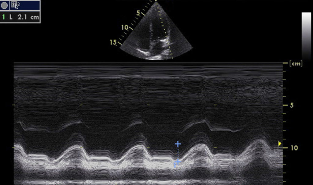
RESEARCH • Feasibility and Discriminatory Value of Tissue Motion Annular Displacement in Sepsis-Induced Cardiomyopathy: A Single-Center Retrospective Observational Study
There is no formalized or consensus definition of SICM, yet frequently left ventricular ejection fraction (LVEF) of less than 50% in septic patients is often considered as indicative of SICM. The mortality among septic patients with SICM is 2 ~ 3 times higher than those without SICM. Echocardiography is considered the most important method for the diagnosis of SICM. In addition to LVEF, mitral annular plane systolic excursion (MAPSE) is another simple method which is obtained by M-mode echocardiography and commonly used to detect LV dysfunction.
….
Right Ventricular Postsystolic Strain Curve Morphology before and after Vasodilator Treatment in Idiopathic Pulmonary Arterial Hypertension
The authors discussed the current guidelines for diagnostic workup of pulmonary hypertension, which include right heart catheterization to establish diagnosis, a transthoracic echocardiography to rule out left heart disease, a ventilation/perfusion lung scan to exclude chronic thromboembolic pulmonary hypertension, a chest computed tomography scan and pulmonary functional tests to exclude parenchymal lung disease, and routine screening of connective tissue disease, hepatitis, and HIV….
ECHOCARDIOGRAPHY AND PULMONARY HYPERTENSION • Echocardiographic Biventricular Coupling Index to Predict Precapillary Pulmonary Hypertension
Right heart catheterization (RHC) remains the gold standard in the diagnostic workup. However, transthoracic echocardiography is currently part of the screening process and is increasingly used in the longitudinal follow-up of patients with PH. Estimated systolic pulmonary artery pressure (sPAP) is recommended to define the probability of PH in symptomatic patients but does not discriminate between pre- and postcapillary PH…
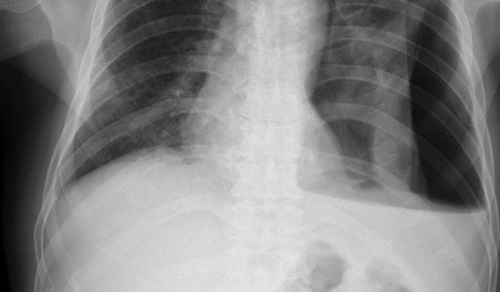
ORIGINAL ARTICLE • Prehospital Portable Ultrasound for Safe and Accurate Prehospital Needle Thoracostomy: A Pilot Educational Study
Since prehospital POCUS is becoming more widely adopted for its diagnostic abilities to aid in decision-making in traumatically injured patients, the authors designed this pilot educational study intervention to train ALS prehospital personnel to use a handheld POCUS to aid in the detection of pneumothorax and apply it during the procedure to improve safety and success…
ORIGINAL ARTICLE • Relationship of the Internal Jugular Vein to the Common Carotid Artery: Implications for Ultrasound-Guided Vascular Access
The internal jugular vein (IJV) is a commonly used cannulation site for central venous access as it is easily accessible due to its superficial anatomical position. The traditional landmark-based technique for IJV cannulation depends on it having a lateral position relative to the common carotid artery (CCA)…
PERICARDIAL FAT AND CARDIAC FUNCTION • Association of Pericardial Fat with Cardiac Structure, Function, and Mechanics: The Multi-Ethnic Study of Atherosclerosis
Among 6,814 participants initially recruited in MESA, the authors included the 3,032 participants who underwent echocardiography at exam 6. At baseline, this cohort was 53% female, with mean age 57 years, and 40% White, 25% Black or African American, 22% Hispanic, and 13% Asian…
CASE STUDY • ‘Hydro-point’ – The Forgotten and Unspoken Entity in Hydropneumothorax
Lung point-of-care ultrasound (POCUS) showed hydro-point, dynamic air–fluid interface (DAFI) sign and a characteristic triple sign, namely barcode sign/stratosphere sign, hydro-point and sinusoidal pattern in right lung. These features are seen in effusive pneumothorax…
RESEARCH • Intermediate-Risk Pulmonary Embolism: Echocardiography Predictors of Clinical Deterioration
Acute pulmonary embolism (PE) can lead to clinical deterioration due to its effect on the right ventricle (RV). Since there are no consistent definitions or assessments of abnormal RV (abnlRV) there is limited data on association between abnlRV and clinical outcomes and prognostic performance…
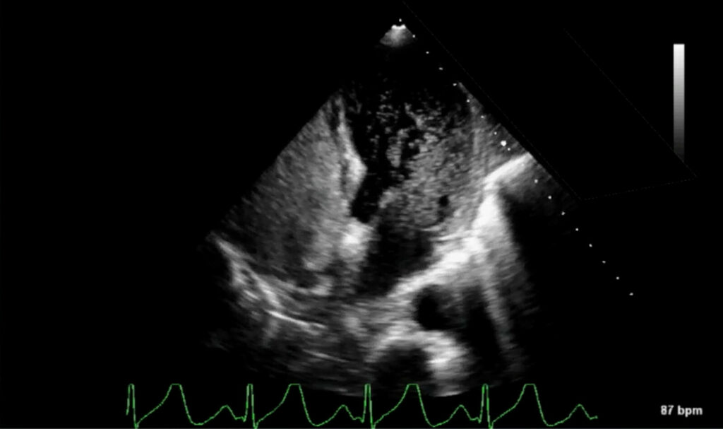
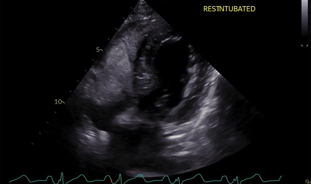
CASE REPORT • Paradoxical Venous Air Embolism Detected with Point-of-Care Ultrasound
Based on ultrasonographic findings the suspected etiology of shock was venous gas embolism with global ventricular systolic dysfunction. Paradoxical embolism was considered based on the presence of bubbles in left chambers during FATE evaluation…
PERICARDIAL FAT AND CARDIAC FUNCTION • Association of Pericardial Fat with Cardiac Structure, Function, and Mechanics: The Multi-Ethnic Study of Atherosclerosis
The authors utilized data from the Multi-Ethnic Study of Atherosclerosis (MESA) to examine the association between pericardial adipose tissue volume by computed tomography (CT) with cardiac structure and function by echocardiography…
KELETAL MUSCLE PERFUSION DURING MECHANICAL CIRCULATORY SUPPORT • Contrast Ultrasound Assessment of Skeletal Muscle Recruitable Perfusion after Permanent Left Ventricular Assist Device Implantation: Implications for Functional Recovery
While diagnosis can be made with computed tomography, magnetic resonance imaging, cardiac catheterization, and echocardiography, only echocardiography which has the advantage of low cost and good safety profile, is convenient for use as a screening tool…
CASE REPORT • The Dance of Death: Cardiac Arrest, Mitral and Tricuspid Valve Prolapses, and Biannular Disjunctions
Despite advances in resuscitation techniques, out-of-hospital cardiac arrests (OHCAs) remain a pressing global health issue with poor outcomes. In this case report, the authors describe a patient with OHCA secondary to ventricular fibrillation that was found to have the classical features of MVP, MAD, TVP, and TAD on multimodality cardiac imaging…
ECHOCARDIOGRAPHY IN CHILDREN • Doppler Echocardiographic Features of Pulmonary Vein Stenosis in Ex-Preterm Children
PVS in children causes significant morbidity and mortality (estimated 3-year survival rate of 43%). While diagnosis can be made with computed tomography, magnetic resonance imaging, cardiac catheterization, and echocardiography, only echocardiography which has the advantage of low cost and good safety profile…
ORIGINAL RESEARCH • Diagnostic Utility of Point of Care Ultrasound in Identifying Cervical Spine Injury in Emergency Settings
CT scan has emerged as a good screening tool in C-spine injury due to its high negative predictive value. However, in a hemodynamically unstable trauma patient, many clinical guidelines do not recommend CT scan, which results in delayed clearance of C-spine in these unstable patients….
SHORT COMMUNICATION • Design and Comparison of a Hybrid to a Traditional In-Person Point-of-Care Ultrasound Course
Point-of-care ultrasound (POCUS) reduces procedural complications, improves diagnostic accuracy, and increases provider and patient satisfaction. In addition, the availability of less expensive ultrasound devices and the increasing accessibility of training programs have facilitated the growth of POCUS to medical students…
BRIEF RESEARCH COMMUNICATIONS • Serial Assessment of Tricuspid Annular Plane Systolic Excursion Is Associated with Death or Lung Transplant in Children with Pulmonary Arterial Hypertension (PAH)
Pulmonary arterial hypertension (PAH) in the pediatric population may be classified as primary/idiopathic or may be associated with congenital heart (CHD) or lung disease. Over time, right ventricular (RV) function determines morbidity and mortality…
PERIOPERATIVE MEDICINE • Pregnancy and Labor Epidural Effects on Gastric Emptying: A Prospective Comparative Study
The goal of this study was to determine and compare the gastric emptying after intake of a standardized light meal in women in labor with epidural analgesia, women in labor without epidural analgesia, pregnant women not in labor, and nonpregnant women using a reliable, noninvasive ultrasound tool…
REVIEW & ANALYSIS • Incidence of Gastrointestinal Bleeding after Transesophageal Echocardiography in Patients with Gastroesophageal Varices: A Systematic Review and Meta-Analysis
Transesophageal echocardiography (TEE) is an important diagnostic procedure that is specifically useful in patients with advanced cirrhosis since this group of patients frequently has cirrhotic cardiomyopathy with varying degrees of heart failure…
CASE STUDY • The Role of Ultrasound in the Diagnosis of Grynfeltt–Lesshaft Lumbar Hernia
Primary lumbar hernia tend to grow over time and start to present symptoms. If undiagnosed by point-of-care ultrasound or if left untreated lumbar hernias could result in death because of the high risk of complications…
ORIGINAL CONTRIBUTION: Early Point-Of-Care Ultrasound Assessment for Medical Patients Reduces Time to Appropriate Treatment: A Pilot Randomized Controlled Trial
Point-of-care ultrasound (POCUS) has proven to be an effective tool that narrows the differential diagnosis among patients presenting with chest pain and dyspnea, which can help achieve the correct diagnosis faster…
IMAGES IN CARDIOVASCULAR ULTRASOUND • A Rare Case of Left Ventricular Noncompaction in LEOPARD Syndrome
Transthoracic echocardiography and cardiac magnetic resonance imaging revealed left ventricular noncompaction* and skin biopsy had no evidence of malignancies. Finally, other disease with hyper-pigmented skin lesions including Addison’s disease, hemochromatosis, and hyperthyroidism, were excluded…
ARTICLE REVIEW • Ultrasound-Enhancing Agents and Associated Adverse Reactions: A Potential Connection to the COVID-19 Vaccines?
The authors report of adverse reactions after administration of Lumason and Definity across three echocardiography laboratories within the University of Pennsylvania Health System (UPHS) and the Medical University of South Carolina (MUSC) from 2019 to 2021…
ORIGINAL ARTICLE • Transcutaneous Laryngeal Ultrasonography: A Reliable, Non-Invasive and Inexpensive Preoperative Method In the Evaluation of Vocal Cords Motility—A Prospective Multicentric Analysis on a Large Series and a Literature Review
Transcutaneous laryngeal ultrasonography (TLUS) has been proposed as a non-invasive and painless indirect examination of vocal cords function as alternative to direct flexible fiberoptic laryngoscopy (FFL). The study assessed TLUS reliability as an alternative method to direct FFL in evaluation vocal folds function for thyroid surgery…
JOURNAL REVIEW • Point of Care, Clinician-Performed Laryngeal Ultrasound and Pediatric Vocal Fold Movement Impairment
An alternate way of assessing vocal fold mobility can be achieved with transcervical laryngeal ultrasound (LUS), which causes fewer changes in physiologic parameters. The goal in this study was to evaluate the use of LUS as a point of care, otolaryngologist-performed assessment of vocal fold movement in the pediatric population…
ORIGINAL RESEARCH • Contrast-enhanced Ultrasound is Useful for the Evaluation of Focal Liver Lesions in Children.
While liver tumors are rare in children, they can pose a diagnostic challenge when suspected. Contrast-enhanced computer tomography (CECT) and contrast enhanced magnetic resonance imaging (CEMRI) are the main imaging modalities in the workup of liver tumors.. …
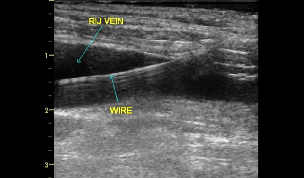
ORIGINAL INVESTIGATION • Internal Jugular Vein Cannulation Using a 3-Dimensional Ultrasound Probe in Patients Undergoing Cardiac Surgery: Comparison Between Biplane View and Short-Axis View
Compared with the traditional anatomic landmark technique, 3-dimensional ultrasound guided IJV cannulation improves patient outcomes by increasing success rates and reducing complications. Both techniques have advantages and disadvantages. …
ORIGINAL INVESTIGATION • Left Atrial Expansion Index for Noninvasive Estimation of Pulmonary Capillary Wedge Pressure: A Cardiac Catheterization Validation Study
RHC, an invasive and potentially risky procedure, is the gold standard technique for accurate hemodynamic assessment and PCWP measurement. However, it is reserved to only highly selected cardiac subgroups of patients. However, it is reserved to only highly selected cardiac subgroups of patients …
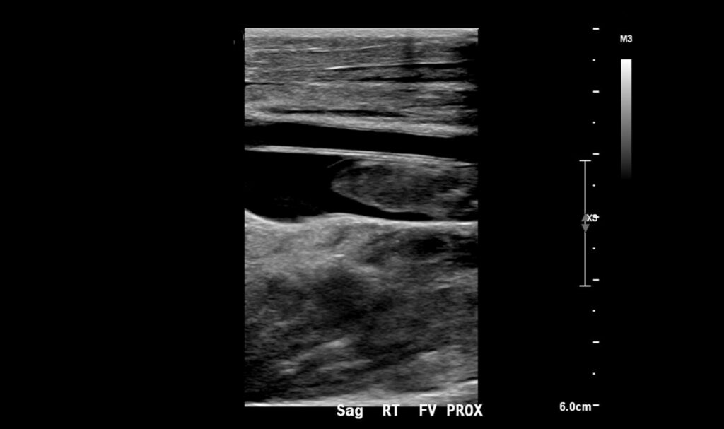
CASE REPORT • Perioperative Point-of-Care Ultrasound for Diagnosis of Acute Lower Extremity Arterial Thrombosis After Total Hip Arthroplasty Revision
In their conclusions, the author comment on the rare complication of acute arterial occlusion after hip arthroplasty, which can lead to devastating outcomes. In the immediate postoperative period, it may be difficult to rely on clinical presentation of acute limb ischemia to provide a timely diagnosis …
RESEARCH ARTICLE • Right Ventricle Early Inflow-Outflow Index May Inform About the Severity of Pneumonia in Patients with COVID-19
Although COVID-19 infection was initially thought to be a disease presenting with viral pneumonia, it may also affect multiple organ systems. The cardiovascular system is one of the most frequently affected systems, and there is an increasing prevalence of cardiovascular complications …
NORMAL VALUES FOR RIGHT VENTRICULAR MEASUREMENTS • Two-Dimensional Echocardiographic Right Ventricular Size and Systolic Function Measurements Stratified by Sex, Age, and Ethnicity: Results of the World Alliance of Societies of Echocardiography Study
Despite the inherent challenges associated with imaging of the anterior, highly asymmetric, crescent-shaped chamber, the right ventricular (RV) size and function is most frequently imaged with echocardiography. A variety of measurements are made to characterize RV chamber size and function …
STATE-OF-THE-ART REVIEW • Ultrasound Imaging of the Abdominal Aorta: A Comprehensive Review
Ultrasound imaging as done in vascular and radiology laboratories typically involves imaging of the entire AA, performed in a fasting state by personnel trained in general and/or vascular ultrasound, and aims primarily at identifying structural abnormalities of the AA …
Ventricular Septal Defect Area by Three-Dimensional Assessment of Shunt Severity in Children
Ventricular septal defect (VSD) size is a determinant of shunt severity in children. When compared to three-dimensional (3D) echocardiography (3DE) and surgical measures, Two-dimensional (2D) echocardiography (2DE) underestimates VSD size …
ORIGINAL ARTICLE • Lung Ultrasound Training and Evaluation for Proficiency Among Physicians in a Low-Resource Setting
Lung ultrasound (LUS) is an effective tool to evaluate patients with dyspnea in the emergency department (ED). While a systematic review of LUS training recommended a three-step approach of teaching theoretical knowledge …
MULTIMODALITY IMAGING DOPPLER DILEMMAS • Pacemaker Lead–Induced Tricuspid Valve Stenosis?
Tricuspid valve (TV) stenosis (TS) is a rare condition and occurs in about 2.4% of cases of primary TV disease. Most commonly, it is caused from rheumatic disease. Endocarditis and large right atrial tumor are other causes of TS …
CLINICAL FOCUS REVIEW • Point-of-care Ultrasound in Cardiac Arrest
Point-of-care ultrasound – an established reliable bedside diagnostic modality for the differentiation of shock – is increasingly used during the management of cardiac arrest. Various resuscitation guidelines advocate for the role of point-of-care ultrasound in cardiac arrest …
VALVULAR HEART DISEASE • Natural History of Moderate Aortic Stenosis with Preserved and Low Ejection Fraction
The authors of this study sought to evaluate if moderate AS is associated with increased mortality in comparison with propensity-matched patients, stratified by ejection fraction (EF), and transaortic gradient, which potentially may justify AV replacement by TAVR …
BRIEF RESEARCH COMMUNICATION • The Impact of Norepinephrine on Myocardial Perfusion in Critical Illness
Myocardial contrast echocardiography (MCE) is a well-validated method of qualitative and quantitative assessment of myocardial perfusion in the non-critically ill. Therefore, the authors’ goal was to investigate whether MCE can be used to assess norepinephrine-related perfusion abnormalities …
RESEARCH • Lung Ultrasound in a Tertiary Intensive Care Unit Population: A Diagnostic Accuracy Study
Etiology of respiratory failure in ICU patients include cardiogenic pulmonary edema (CPE), acute respiratory distress syndrome (ARDS), atelectasis, and pneumonia . Chest X-ray and thoracic computed tomography (CT) are still the most common diagnostic modalities of ICU patients with respiratory failure …
ORIGINAL ARTICLE • Results from a Spanish National Survey on the Application of Ultrasound in Pulmonology Services
While ultrasound use initially started for guidance during pleural procedures, over time it has proven useful in the assessment of the lung parenchyma, diaphragm, and chest wall. Therefore, it has become a basic first-line tool to establish a differential diagnosis in patients with dyspnea…
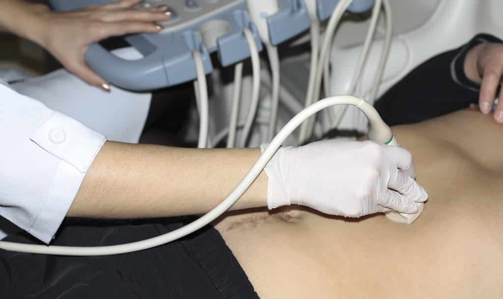
EDUCATIONAL AND CLINICAL PRACTICE ORIGINAL RESEARCH • Bedside Abdominal Ultrasound in Evaluating Nasogastric Tube Placement
Nasogastric tube (NGT) is commonly used in daily clinical practice for a variety of indications such as administration of enteral nutrition and medications, gastric decompression, irrigation, and diagnostic procedures. Chest radiograph has several disadvantages such as a source of irradiation, time-consuming, and it may not always be definitive.
CASE REPORT • Novel Role of Focused Airway Ultrasound in Early Airway Assessment of Suspected Laryngeal Trauma.
Laryngeal injuries, which are rare, are often undiagnosed in the initial evaluation of the trauma patient. Delayed recognition and intervention may lead to serious complications, including death due to upper airway obstruction …
BRIEF RESEARCH COMMUNICATIONS • Improved Thrombus Assessment by Transesophageal Echocardiography: The DOLOP (Detection of Left Atrial Appendage Thrombosis Utilizing Optison) Study
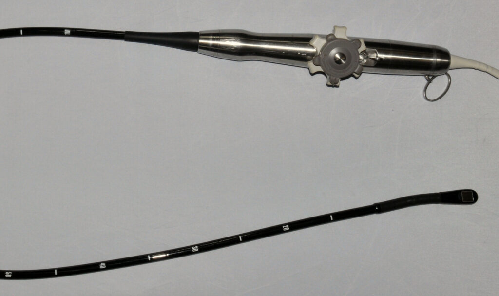
Transesophageal echocardiography (TEE) is an essential clinical tool to assess for thrombus in the left atrial appendage (LAA) prior to cardioversion or ablation in the setting of atrial fibrillation (AF) or flutter …
EDUCATION AND CLINICAL PRACTICE: ORIGINAL RESEARCH • Methodologic Quality of Guidelines for Training or Competence Processes for Basic Point-of-Care Echocardiography in Critical Care – A Systematic Review of the Literature
Point-of-care ultrasound (PoCUS) is defined as a diagnostic or procedural guidance ultrasound performed by clinicians to help guide patient management. Current pathways for PoCUS training, education and competence rely on expert guidelines…
RESEARCH REPORT • Gastric Emptying Time of Two Different Quantities of Clear Fluids in Children: A Double Blinded Randomized Controlled Study
Perioperative pulmonary aspiration in children is infrequent and is more likely to occur in emergency situations than in elective surgeries. Gastric ultrasound in pediatric patients could help identify children with significant gastric volumes before anesthesia…
CORRESPONDENCE • Transthoracic Utilization of the Transesophageal Echocardiography Probe—A Novel Window to Non-Invasive Hemodynamic Monitoring for the Pediatric Anesthesiologist
The precordial stethoscope has been the universal standard for continuous cardiorespiratory monitoring during pediatric anesthesia. This long-standing standard has been replaced with transesophageal echocardiography (TEE)…
Clinical Investigations • Simple Two-Dimensional Echocardiographic Scoring System for the Estimation of Left Ventricular Filling Pressure
A noninvasive method of LVFP estimation is necessary in the clinical management of patients with HF. Echocardiography is the first-line tool both for the diagnosis and follow-up of patients with HF…
REPORTS OF ORIGINAL INVESTIGATION • Ultrasound Evaluation of Inferior Vena Cava Compression in Tilted and Supine term Parturients
The authors of this study demonstrated that ultrasound imaging of the IVC is easy to acquire and can detect IVCCI changes in the supine versus tilted position in term parturients…
ORIGINAL ARTICLE • A Retrospective Evaluation of Point of Care Ultrasound for Acute Cholecystitis in a Tertiary Academic Hospital Setting
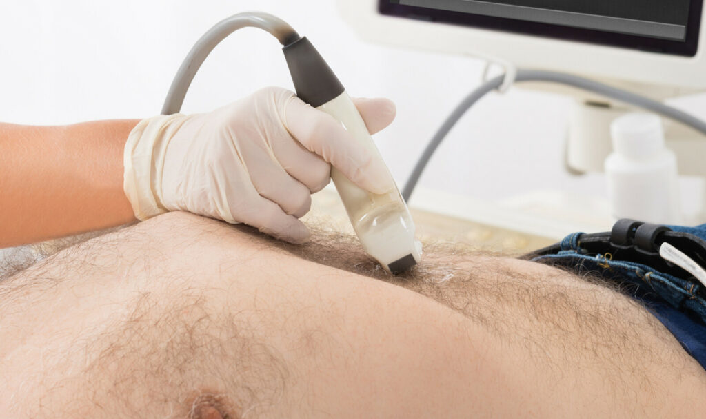
To diagnose acute cholecystitis, clinicians in the emergency department (ED) routinely order radiology-performed ultrasound (RADUS) of the gallbladder to “rule out” acute cholecystitis…
Assessment of Diaphragmatic Function by Ultrasonography: Current Approach and Perspectives
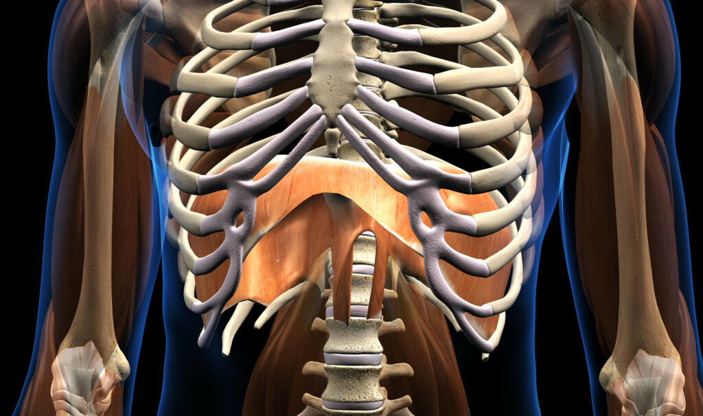
Ultrasonography (US) for the assessment of diaphragmatic function has been reported for 25 years. Several methods have been proposed to evaluate diaphragmatic function…
CLINICAL INVESTIGATION • A Doppler Echocardiographic Pulmonary Flow Marker of Massive or Submassive Acute Pulmonary Embolus
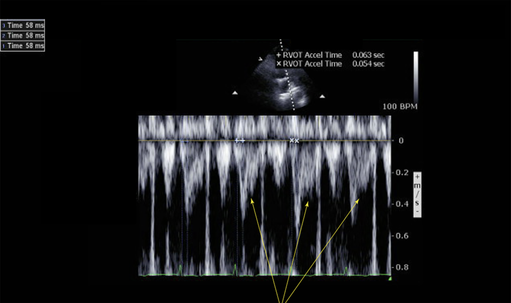
The role of echocardiography in the workup of suspected PE has historically been supportive and primarily reserved for the detection of RV strain or dysfunction in unstable patients….
GENERAL MEDICINE/SYSTEMATIC REVIEW SNAPSHOT • Is Chest Ultrasonography Superior to Supine Chest Radiography in Identifying Pneumothorax in Emergency Department Trauma Patients?
In this clinical synopsis, the authors included 9 studies comprising 1,271 patients with 410 pneumothoraces from an initial 2,293 references. The median prevalence of traumatic pneumothorax was 30% (range 21% to 52%). The overall sensitivity of chest ultrasonography was 0.91…
ECHOCARDIOGRAPHY IN COVID-19 • The Predictive Role of Combined Cardiac and Lung Ultrasound in Coronavirus Disease 2019
Despite the recognition of the prognostic significance and clinical implications of the cardiac complications of coronavirus disease 2019 (COVID-19), both the European Association of Cardiovascular Imaging and the American Society of Echocardiography recommend limiting the echocardiographic assessment of patients with COVID-19…
ORIGINAL RESEARCH • Pneumothorax Diagnosis with Lung Sliding Quantification by Speckle Tracking: A Prospective Multicentric Observational Study
Lung ultrasound is commonly used for the diagnosis of pneumothorax, when the absence of pleural sliding, B-lines and lung pulse, or by the presence of a lung point, has been recommended to make the ultrasound diagnosis of pneumothorax…
PERIOPERATIVE MEDICINE • Ultrasound Evaluation of Gastric Emptying Time in Healthy Term Neonates after Formula Feeding
The current American Society of Anesthesiologists (ASA) fasting guideline for formula-fed infants in the periprocedural setting is 6 h before elective procedures requiring general anesthesia, regional anesthesia, or sedation/analgesia….
QUALITY IMPROVEMENT REPORT • Improving the Documentation Quality of Point-of-Care Ultrasound Scans in the Emergency Department
Point-of-care ultrasound scan (POCUS) has been incorporated a as a mandatory element of the Royal College of Emergency Medicine (RCEM)since 2010. All trainees must be signed off as competent in the four CORE clinical applications…
HEAD-TO-HEAD • Should Point-of-Care Ultrasonography Replace Stethoscopes in Acute Respiratory Failure?
When it comes to diagnosing the cause of acute respiratory failure, the current standard is to use the stethoscope and chest radiography—two technologies with a combined age of over 300 years….
ORIGINAL ARTICLE | Scoping Review of Implementing a Longitudinal Curriculum in Undergraduate Medical Education: The Wake Forest Experience.
The integration of hands-on ultrasound experience has become a highly desirable component for undergraduate medical education (UGME) curricula throughout the United States. Several schools have implemented a student-performed ultrasound experience…
ORIGINAL ARTICLE | Case series: Point-of-Care Ultrasound Conducted by Medical Students During their First Clinical Rotation Changes Patients’ Primary Diagnosis and Management
The authors point out that we practice medicine in a transitional time where POCUS has entered the curriculum of some medical schools, but many senior and experienced physicians do not hold this new bedside capability…
ORIGINAL RESEARCH | Emergency Medicine Advanced Ultrasound Service: A New Paradigm
The use of point-of-care US (POCUS) has become well established in emergency medicine (EM), largely due to social and economic pressures to provide rapid assessment and transit through the emergency department (ED) and the desire for improved diagnostic and procedural ability in acute care settings…
REVIEW | Effect of Intermittent High–Mechanical Index Impulses on Left Ventricular Strain
Endocardial border resolution can be improved by using Intermittent high–mechanical index (MI) impulses during ultrasound enhancing agent (UEA) infusion. In animal models, these high-MI impulses have been shown to also release adenosine triphosphate (ATP) for sustained periods of time…
MITRALVALVE DISEASE | Mitral Valve Prolapse Patients with Less than Moderate Mitral Regurgitation Exhibit Early Cardiac Chamber Remodeling
Mitral valve prolapse (MVP), which has a prevalence of 2.4% in the general population, is the most common etiology of mitral regurgitation (MR). The natural history of MVP is diverse, ranging from slow progression of mild MR degrees, to rapid progression to severe MR requiring cardiac surgery….
REVIEW | A Systematic Review of the Cost-Effectiveness of Ultrasound in Emergency Care Settings
Ultrasound (US) is safe and effective imaging modality for the rapid diagnosis and management of emergency conditions. Technological advances and the increasing availability of imaging modalities led to expansion of the use of clinical US. It is assumed that US is cost-effective in emergency care settings….
ORIGINAL INVESTIGATION | Utility of a Deep-Learning Algorithm to Guide Novices to Acquire Echocardiograms for Limited Diagnostic Use
The believe that this AI guidance algorithm represents a step forward in the interaction of AI with medical imaging and may allow for extension of ultrasonography to settings that ordinarily do not have access…
ORIGINAL ARTICLE | The Reproducibility of Measuring Maximum Abdominal Aortic Aneurysm Diameter from Ultrasound Images
Findings suggest that abdominal ultrasound measurements of AAA size should be performed in the anterior–posterior plane and ideally compared between different time periods by the same observer……
CLINICAL STUDY | Carotid Ultrasound in Assessing Fluid Responsiveness in Patients with Hypotension and Suspected Sepsis
Since over-and under-resuscitation are associated with adverse outcome, it is important to identify patients who would benefit from additional fluid administration after initial intravenous fluid (IVF) bolus…..
A DIAGNOSTIC ACCURACY STUDY | Auscultation Versus Point-of-care Ultrasound to Determine Endotracheal Versus Bronchial Intubation
While it is relatively straightforward to discriminate tracheal intubation from esophageal intubation, identifying correct location of the endotracheal tube (ETT) within the trachea remains challenging….
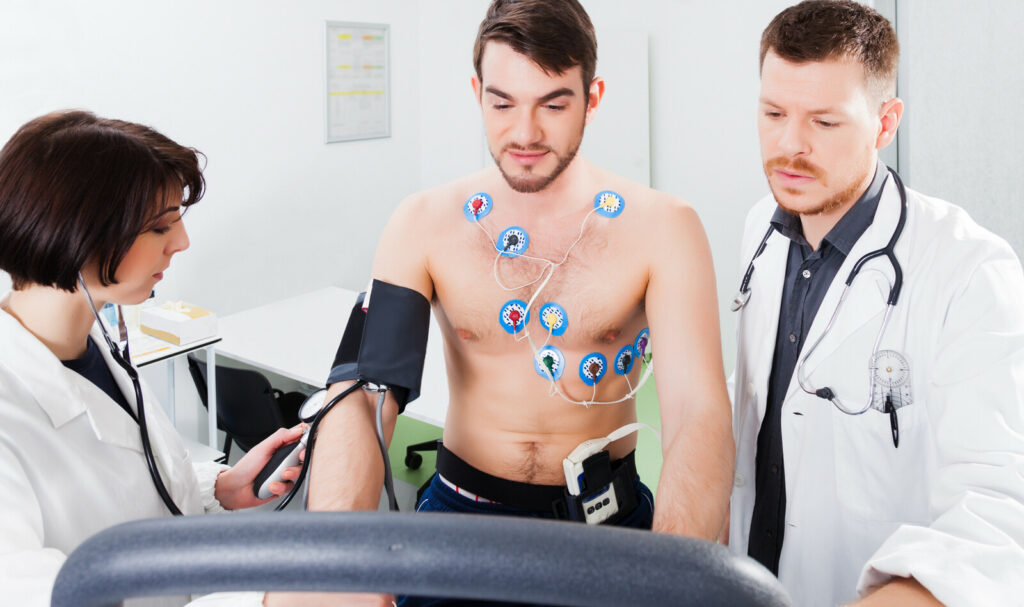
STATE OF THE ART REVIEW | Usefulness of Stress Echocardiography in the Management of Patients Treated with Anticancer Drugs
Stress echocardiography has a pivotal role in a timely diagnosis of CAD. The atherosclerotic process can be accelerated by both chemotherapy and chest irradiation in patients with cancer, even several years after treatment completion….
ORIGINAL CLINICAL RESEARCH REPORT | Gastric Ultrasound Assessing Gastric Emptying of Preoperative Carbohydrate Drinks: A Randomized Controlled Noninferiority Study
Gastric ultrasound provides quantitative and qualitative information about nature of gastric contents before anesthesia. It is a useful tool for assessing risk of pulmonary aspiration, which has altered clinical management during anesthesia…
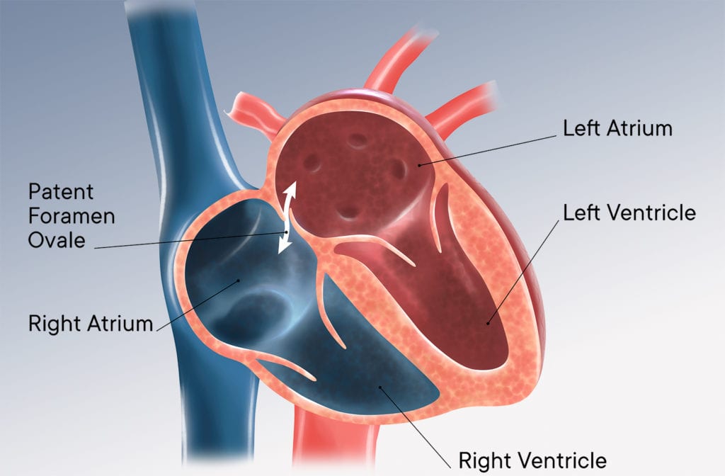
BRIEF RESEARCH COMMUNICATIONS | Efficacy of Saline Contrast Transthoracic Echocardiography for Identifying High-Risk Patent Foramen Ovale
The authors concluded that grade 4 on saline contrast TTE can be valuable for identifying high-risk PFO associated with CS. Saline contrast TTE may be effective to stratify patients for performing. …
ORIGINAL ARTICLE | Teaching Limited Compression Ultrasound to General Practitioners Reduces Referrals of Suspected DVT to a Hospital: a Retrospective Cross-Sectional Study
The authors concluded that teaching LCUS protocol to GPs substantially reduced referrals to the hospital due to a suspected DVT. Importantly, it did not seem to result in an increasing number of pulmonary embolisms…
Systematic Review Article: Intraoperative Focused Cardiac Ultrasound for Assessment of Hypotension: A Systematic Review
The use of focused cardiac ultrasound (FoCUS), which can be performed by non-cardiologists with proper training for cardiovascular evaluation, has gained acceptance in emergency department (ED) & intensive care units…
Research Letter: Lung Ultrasound Can Predict Response to the Prone Position in Awake Non-Intubated Patients with COVID-19 Associated Acute Respiratory Distress Syndrome.
Prone positioning (PP) is often used in acute respiratory distress syndrome (ARDS). While studies have demonstrated positive effects on oxygenation in awake and non-intubated patients, it isn’t effective in every case…
CASE REPORT | Native Mitral Valve Infective Endocarditis From Flossing: A Case Report and Emergency Department Management
Infective endocarditis (IE) is often classified into left (mitral and aortic), right (tricuspid and pulmonic), as well as by the nature of the affected valve (native or prosthetic). 40% of these cases affect the mitral valve. …
STATE OF THE ART REVIEW | Agitated Saline Contrast Echocardiography in the Identification of Intra- and Extracardiac Shunts: Connecting the Dots
Cardiologists throughout the world often perform and interpret echocardiographic studies using agitated saline as a contrast agent. Also known as ‘‘bubble studies’’, these studies can provide valuable information…
ECHOCARDIOGRAPHY IN CHILDREN | Longitudinal Variation in Presence and Severity of Cardiac Valve Regurgitation in Healthy Children
In both normal adults and pediatric populations, trivial or mild valvular regurgitation (VR) is a common finding. However, in a specific individual, such finding may raise the concern whether this is a physiologic or pathologic finding…
BRIEF RESEARCH COMMUNICATION – COVID 19 | Mitral Annular Plane Systolic Excursion: An Early Marker of Mortality in Severe COVID-19
Respiratory failure is one of the major causes of death in hospitalized patients with COVID-19. Right ventricular (RV) dilation and reduced RV longitudinal strain have been shown to be markers of poor outcome in this disease…
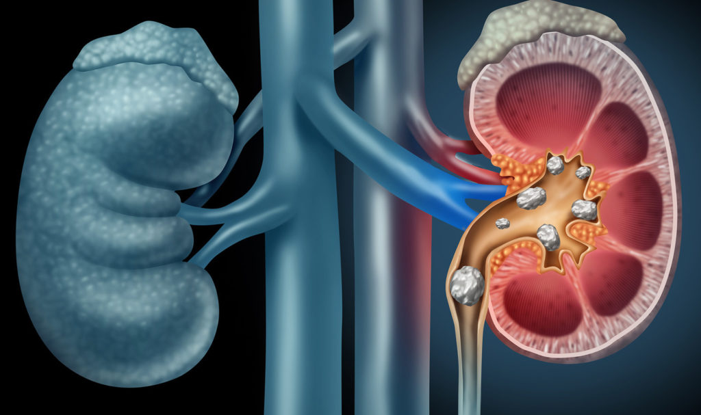
Original Article: Point-of-Care Ultrasound for the Detection of Hydronephrosis in Emergency Department Patients with Suspected Renal Colic.
Renal colic is a common presentation in the emergency department (ED), leading to significant utilization of resources, including advanced imaging as well as direct and indirect costs related to patient care and lost work time…
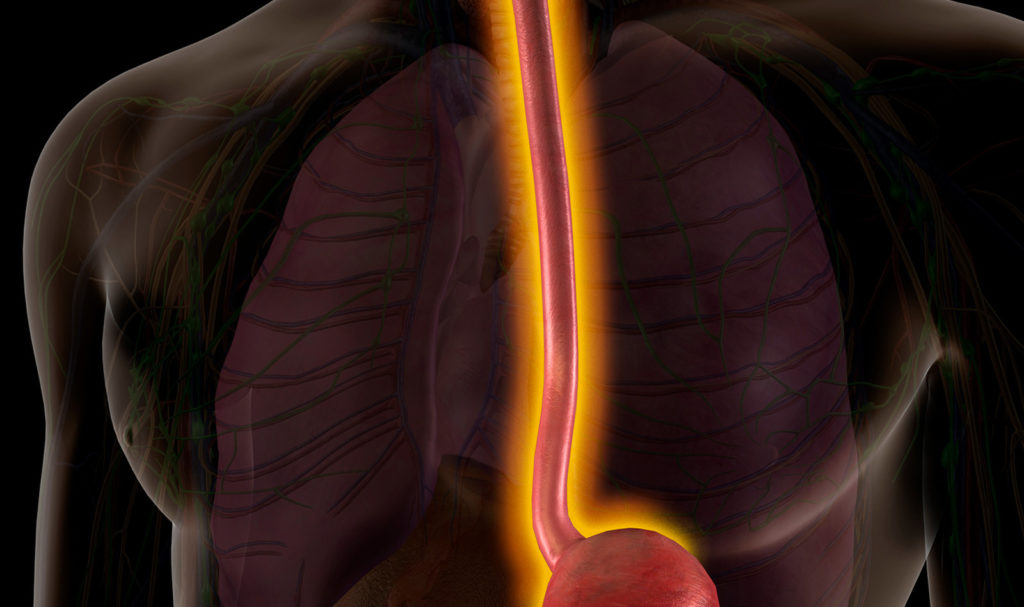
Aortic Awareness Case Review: Esophageal Perforation with Transesophageal Echocardiography in an Elderly Patient
While uncommon, transesophageal echocardiography (TEE) can lead to esophageal perforation which, in rare occasion, can even lead to death. The authors present a case of iatrogenic esophageal perforation from TEE…
Review: Role of Point-of-Care Ultrasound During the COVID-19 Pandemic: Our Recommendations in the Management of Dialytic Patients
LUS is well validated in evaluation of several respiratory conditions. LUS is an image method that classically allows the evaluation of the pulmonary periphery, which is helpful in assessing COVID-19….
Ultrasound-Guided Peripheral Vascular Catheterization in Pediatric Patients: A Narrative Review
In children, the utility of ultrasound-guided PVC placement has been demonstrated in numerous studies, showing improved success rate, procedure time, number of attempts, and number of complications…
Original Article: Unanticipated Critical Findings on Echocardiography in Septic Patients
Use of echocardiography to manage patients with sepsis has become routine. Transthoracic echocardiogram (TTE) now considered appropriate in management of patients in shock or where TTE is likely to change management…
Opinion Paper: Lung Ultrasound in a COVID Pandemic – Choosing Wisely
This paper reviews four important questions that should be answered by each provider, which will help the provider customize lung ultrasound provision: Why, what, who, and when?
Letter: Optic Nerve Sheath Diameter Guided Detection of Sepsis-Associated Encephalopathy
This letter was written in response to the research letter discussing the measurement of ultrasonography guided optic nerve sheath diameter (ONSD) in sepsis associated encephalopathy (SAE) and its correlation with outcome…
Research Letter: Bedside Ultrasound Measurement of Optic Nerve Sheath Diameter in Patients with Sepsis: A Prospective Observational Study
Sepsis-associated encephalopathy (SAE) is a serious condition that can lead to cognitive impairment, sleep disturbances, delirium and even coma. One of the complications of SAE is brain edema leading to high intracranial pressure…
Clinical Investigation: Hypertrophic Cardiomyopathy and Contrast Echocardiography
Hypertrophic cardiomyopathy (HCM) is marked by left ventricular hypertrophy. Left ventricular wall thickness (LVWT) is a key element in the diagnostic workup and prognostic assessment of HCM…
Systematic Review & Meta‐Analysis: Is Contrast-Enhanced Ultrasound (CEUS) Superior to Computed Tomography Angiography (CTA) in Detection of Endoleaks in Post-EVAR Patients?
The author concluded contrast-enhanced ultrasound scan has a higher sensitivity and comparable specificity to computed tomography angiography for detection of endoleaks post-EVAR…
Original Investigation: Right Ventricular Global Longitudinal Strain and Outcomes in Heart Failure with Preserved Ejection Fraction
Studies evaluated prognostic impact of right ventricular dysfunction (RVD) in patients with HF with preserved ejection fraction (HFpEF) found patients with RVD had a worse prognosis…
Clinical Research: Echocardiographic Evaluation of Cardiac Recovery After Refractory Out-of-Hospital Cardiac Arrest
While several techniques have been employed, there is no consensus as to whether unloading strategies should be routinely employed to reduce LV wall stress…
Original Investigation: Incidence of Stress Cardiomyopathy During the Coronavirus Disease 2019 Pandemic
Recent reports suggest that the incidence of stress cardiomyopathy (also known as Takotsubo cardiomyopathy) may have gone up during the COVID-19 crisis.
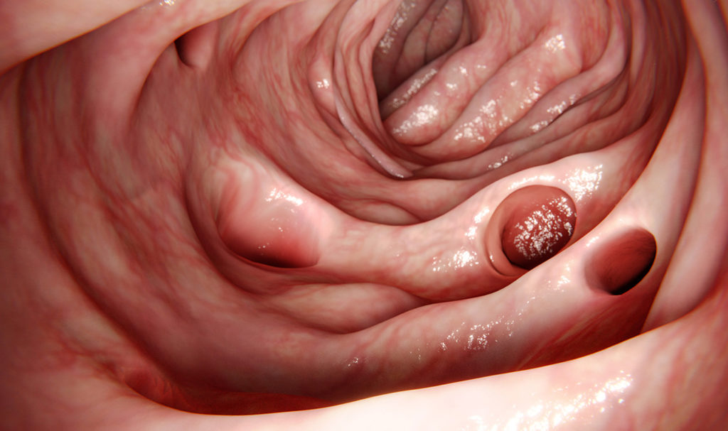
Research Analysis: A Prospective Evaluation of Point-of-Care Ultrasonographic Diagnosis of Diverticulitis in the Emergency Department
Diverticulitis often presents with a constellation of non-specific symptoms, including diarrhea, fever, and abdominal pain, making it challenging to diagnose…
Review: A Systematic Review and Pooled Analysis of CPR-Associated Cardiovascular and Thoracic Injuries
CPR-associated thoracic injuries in setting of manual chest compressions or circulatory adjuncts using active compression–decompression technology is not well characterized…
Review: Recommendations for Core Critical Care Ultrasound Competencies as a Part of Specialist Training
Critical care ultrasound (CCUS) has become an integral part of intensive care practice but only few countries have managed to operationalize this guidance…
Clinical Research: Tissue Doppler Imaging of the Diaphragm in Healthy Subjects and Critically Ill Patients
Ultrasonography has been used to evaluate diaphragmatic motion characteristics, with derived measurements relating to outcome of weaning from ventilation…
Case Report: Stab Wound of the Superficial Femoral Artery Early Diagnosed by Point-of-Care Doppler Ultrasound
Point-of-care Doppler ultrasound was performed and demonstrated a large hypoechoic, slightly pulsating hematoma in the subfascial compartment…
Prospective International Survey: Global Evaluation of Echocardiography in Patients with COVID-19
Accumulating data suggest that COVID-19 can cause a wide range of cardiac conditions that include acute myocardial infarction, myocarditis, and stress cardiomyopathy.
Study of Ethics & Law: Analysis of Lawsuits Related to Diagnostic Errors from Point-of-Care Ultrasound in Internal Medicine, Pediatrics, Family Medicine and Critical Care in the USA.
As adoption of this Point-of-Care ultrasound continues to increase in internal medicine, cardiac care, pediatrics and more, there are some concerns about potential for lawsuits related to its use in clinical practice.
Clinical Investigations: Systematic Assessment of the Impact of Ultrasound-Enhancing Agents upon Measurements of Cardiac Size and Function by Echocardiography
Ultrasound-enhancing contrast agent (CON) is now used routinely to improve both endocardial definition and accurate measurements in patients with suboptimal image quality.
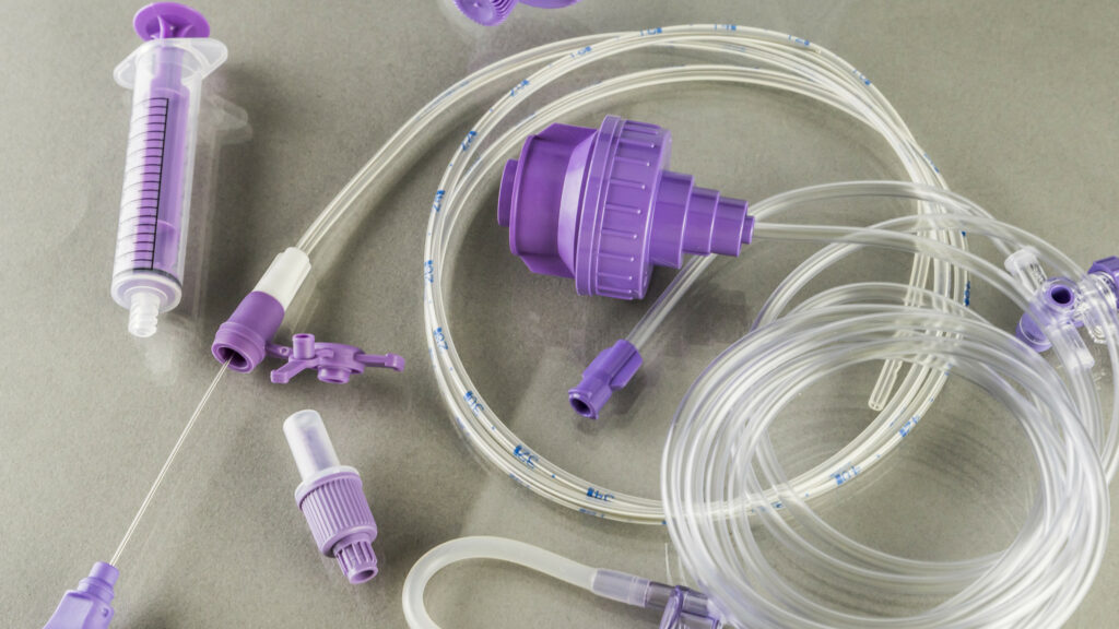
Case Report: Pediatric Case of Successful Point-of-Care Ultrasound-Guided Nasogastric Tube Placement
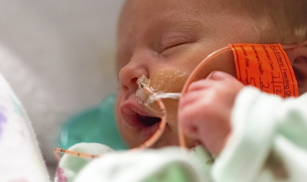
Nasogastric tube (NGT) placement is commonly performed in pediatric emergency care for gastric decompression, enteral feeding, and drug administration, and has the potential to cause serious complications.
Case Report: Point-of-Care Ultrasound in the Diagnosis of Neonatal Cerebral Sinovenous Thrombosis
This is a case of a 27-day-old male neonate who was admitted to the pediatric ward with acute respiratory syncytial virus-bronchiolitis.
Research Letter: A longitudinal cohort of stress cardiomyopathy assessed with speckletracking echocardiography after moderate to severe traumatic brain injury.
When assessed with speckle-tracking echography, stress cardiomyopathy is seen in about a third of patients with subarachnoid hemorrhage (SAH).
Correspondence: Contrast-enhanced ultrasound (CEUS) of the lung reveals multiple areas of microthrombi in a COVID-19 patient
The correspondence letter describes contrast-enhanced ultrasound (CEUS) of the lung, and follows a different letter in Intensive Care Medicine (April 23, 2020) questioning whether areas of subpleural consolidation are indicative of segmental pulmonary embolus.
Research: Regional right ventricular longitudinal systolic strain for detection of severely impaired right ventricular performance in pulmonary hypertension
Right ventricular (RV) function is a key determinant of outcome in patients with pulmonary hypertension (PH), which is a life- threatening disease leading to RV dilatation and failure. Hemodynamics assessed using Invasive approach of right heart catheterization (RHC) provides important information of RV function and prognosis.
Review: Point-of-care ultrasound in pregnancy: gastric, airway, neuraxial, cardiorespiratory.
This is a review article that looks into the use of PoCUS in the anesthetic management of the obstetric patient. The article reviews the use of ultrasound for assessment of aspiration risk, airway management, placement of neuraxial blocks and the diagnosis and follow-up of cardiorespiratory dysfunction.
Review: Rolling a trauma patient onto the right side increases sensitivity of FAST examination.
Shortening time for hemoperitoneum diagnosis may improve morbidity and mortality if time from diagnosis to operating room is shortened. Therefore, techniques that increases the sensitivity of FAST exam may lead to reduced morbidity & mortality.
Analysis: Self-learning of point-of-care cardiac ultrasound — Can medical students teach themselves?
Training of PoCUS is challenging, time-consuming and resource-intensive. This study evaluated whether training process can be simplified by allowing medical students to self-train with web-based cardiac ultrasound software.
Abstract: Echocardiography – POCUS Training Recommendations
Recommendations for Echocardiography Laboratories Participating in Cardiac Point of Care Cardiac Ultrasound (POCUS) and Critical Care Echocardiography Training: Report from the American Society of Echocardiography.
Abstract: Echocardiography – Diagnosis of PFO
Importance of Abdominal Compression Valsalva Maneuver and Microbubble Grading In Contrast Transthoracic Echocardiography for Detecting Patent Foramen Ovale.
Abstract: Pediatric POCUS: Skull Fractures Detection
The utility of Ultrasound in detecting skull fractures after pediatric blunt head trauma. Systematic review and meta-analysis.
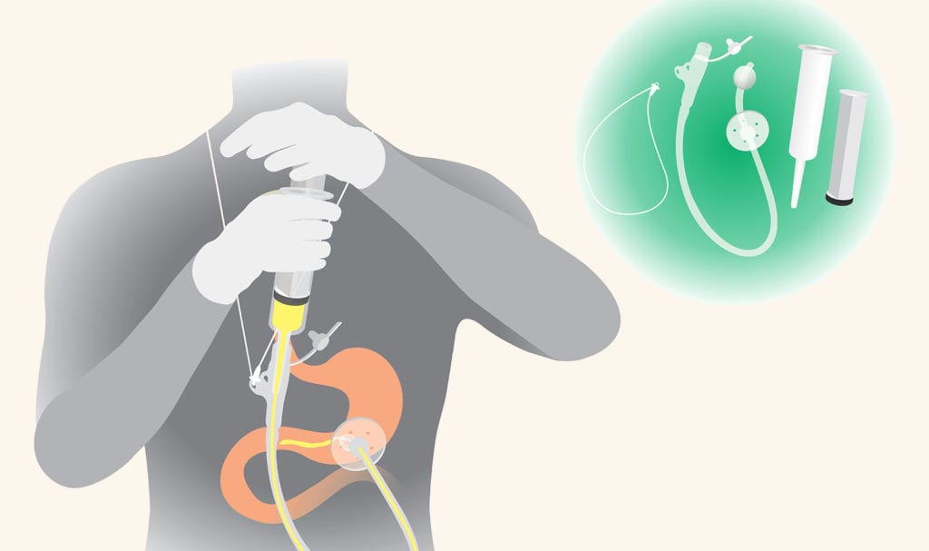
Abstract: Pediatric POCUS: G-Tube Confirmation
Point-of-care ultrasound for confirmation of gastrostomy tube replacement in the pediatric emergency department
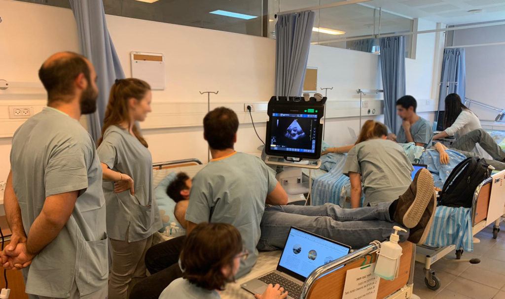
eMedical Academy Publishes New Case Study Detailing Partnership with Winfocus-Switzerland to Train Cohort of Medical Students on Point of Care Ultrasound.
eMedical Academy online ultrasound school for Point-of-Care ultrasound continuing education recently published a new case study on their website. The case study explores how Winfocus-Switzerland incorporated virtual ultrasound courses from eMedical Academy to train its medical students…
eMedical Academy Publishes New Case Study Discussing Partnership with the Department of Anesthesia in the Children’s Hospital of Boston
eMedical Academy online ultrasound school for Point-of-Care ultrasound continuing education recently published a new case study on their website. The case study explores how the Department of Anesthesia in Boston’s Children’s Hospital decided to use virtual ultrasound courses from eMedical Academy to train staff in multiple locations to meet departmental objectives…
eMedical Academy Publishes New Case Study Outlining Partnership to Train Ultrasound Students of Technion Medical School’s Mission in Uganda
eMedical Academy online ultrasound school for Point-of-Care ultrasound continuing education recently published a new case study on their website. The case study explores how Technion Medical School incorporated virtual ultrasound courses from eMedical Academy to train its Mission in Uganda medical students…
eMedical Academy Publishes New Case Study Sharing Details of Partnership with Ben-Gurion University of the Negev in Beersheba, Israel
eMedical Academy online ultrasound school for Point-of-Care ultrasound continuing education recently published a new case study on their website. The case study explores how the Ben-Gurion University of the Negev in Beer Sheva, Israel, incorporated the virtual ultrasound courses from eMedical Academy into the university’s medical school curriculum…
 English
 Español

