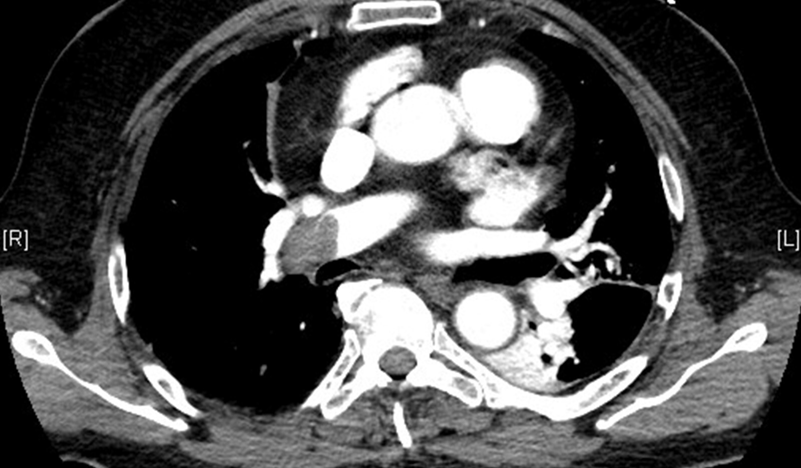
RESEARCH • Intermediate-Risk Pulmonary Embolism: Echocardiography Predictors of Clinical Deterioration
Source: Critical Care (2022) 26:160
INTRODUCTION
Acute pulmonary embolism (PE) can lead to clinical deterioration due to its effect on the right ventricle (RV). Since there are no consistent definitions or assessments of abnormal RV (abnlRV) there is limited data on association between abnlRV and clinical outcomes and prognostic performance.
Although TTE can qualitatively and quantitatively assess RV size and systolic function, there is no consensus on which of many different TTE metrics define the standard for establishing the presence of clinically impactful RV abnormality. In addition, it is unlikely the different laboratory and image assessments or definitions of RV abnormality can be used interchangeably.
Although the American Society of Echocardiography (ASE) provides guidelines for RV chamber size and systolic function measurements for adults without cardiac or pulmonary disease, there are no disease-specific reference ranges for RV to grade disease severity. Physicians using different risk stratification models for determining abnlRV may arrive at different disposition decisions, clinical management considerations, and intervention decisions on any given PE patient.
RV size or systolic function is included as dichotomous predictors in some PE triaging strategies (e.g., Bova score and European Society of Cardiology guidelines). However, those strategies do not link the severity of RV abnormalities with their associated probability of immediate clinical deterioration.
The goal of this study was to identify TTE metrics that distinguish intermediate-risk PE patients at risk for clinical deterioration from those who do not experience death or clinical deterioration within 5 days.
METHODS
Study Design and Setting
This was a prospective observational study within an integrated healthcare system that uses a multidisciplinary approach to the identification of intermediate-and high-risk PE patients, which triggers “Code PE” notifications to clinicians on duty.
Selection of Participants
The authors used a prospective cohort design to identify and study adult patients ( ≥ 18 years) presenting to a participating ED, who had: (1) acute symptomatic PE as the primary ED diagnosis (by positive CT, high-probability ventilation/perfusion nuclear imaging, or point-of-care TTE findings highly suspicious of PE [e.g., RV dilatation on point-of-care, DVT findings]), (2) intermediate-risk PE classification defined as normotensive, with suspected or confirmed RV abnormalities, and (3) comprehensive TTE with RVfocused measurements completed within 24 hours of PE diagnosis. PE risk stratification was based on review of vital signs, CT findings, natriuretic peptide, troponin, and bedside TTE findings at presentation.
To create a quasi-control group, the authors randomly selected 25 PE patients from a previously reported PE registry, who had comprehensive TTE performed and were not classified as intermediate or high risk.
Outcome Measures
The primary outcome was a composite of death, circulatory or respiratory deterioration (including cardiac arrest, respiratory failure, new unstable dysrhythmia, sustained hypotension treated with either fluid or adrenergic agents), or escalated PE intervention within 5 days of PE diagnosis.
The secondary outcome extended the time frame to 30 days after PE and included all components of the primary outcome in addition to major bleeding episodes, recurrence of venous thromboembolism (VTE), and subsequent hospitalization.
RESULTS
Out of 501 Code PE patients that were screened, 306 intermediate-risk patients met criteria for complete analysis, 303 (98%) of whom had CT with RV/LV ratios reported. Of the remaining 3 patients without CT, two (0.6%) had high probability ventilation/perfusion mismatch findings, and two had RV dilatation by point-of-care TTE (one with coexisting lower extremity DVT and one patient had clot in transit on point-of-care TTE as the confirmatory study). All 306 PE patients had troponin measurements and 191 (62.4%) had troponin elevations. Of the 306, 293 patients (95.7%) had BNP measurements, 192 of whom (65.5%) had BNP elevation. Of the 303 with CT, 237 (78.2%) had CT with RV/LV ratio ≥ 1.0.
Within 5 days, 115 (37.6%) patients experienced one or more clinical deterioration events or required an emergent in-hospital intervention. There were 3 (1.0%) deaths; 66 patients (21.6%) had one or more escalated PE interventions; 43 (14.1%) had symptomatic sustained hypotension addressed with intravenous fluid boluses; 31 (10.1%) had respiratory failure; 31 (10.1%) had new dysrhythmia requiring treatment; 20 (6.5%) had sustained hypotension treated with adrenergic agents; and 12 patients (3.3%) had cardiac arrest. There were no clinical deterioration events among the quasi-control group.
Based on the analyzed data, a TAPSE < 1.83 cm results in the optimal threshold for predicting higher risk of clinical deterioration based on TAPSE alone, such that patients with TAPSE < 1.83 had 3.35 times greater odds of clinical deterioration than those with TAPSE ≥ 1.83 cm. Using this threshold for prediction of clinical deterioration results in 57% sensitivity, 71% specificity, 52% PPV, and 76% NPV.
Logistic Regression
The significant abnlRV variables from the best fitting abnlRV model were troponin, BNP, tricuspid regurgitant peak velocity, RV/LV basal width ratio, and TAPSE. Based on the authors’ analysis, significant independent predictors were (estimated odds ratios, 95% confidence intervals, and p values, respectively): transient hypotension 6.1 (2.2, 18.9), highest heart rate within the initial 3 h 1.02 (1.00, 1.03), highest respiratory rate within initial 3 h 1.02 (1.00, 1.04), and RV/LV ratio 1.29 (1.14, 1.47).
Among the abnlRV variables, BNP (p = 0.008) and RV/LV basal width ratio (p < 0.001) were statistically significantly related to increased odds of clinical deterioration, while tricuspid regurgitant peak velocity was marginally significant (p = 0.062). TAPSE and elevated troponin were not independent predictors and were dropped from the model.
CONCLUSIONS
The authors concluded that intermediate-risk PE patients with 5-day clinical deterioration had significantly increased BNP and troponin, increased RV/LV ratio and RV basal diameter, and decreased LV basal diameter and RV systolic function (TAPSE and S’) measurements.
The authors further commented that while further work is needed to better understand the relationships between these variables and their independent prognostic ability, these results suggest that identified variables in this study (e.g., TAPSE, RV/LV basal width ratio, BNP) may provide simple, but useful criteria for assessing patient risk of 5-day clinical deterioration in intermediate-risk PE patients.
 English
 Español

