ORIGINAL CONTRIBUTION: Early Point-Of-Care Ultrasound Assessment for Medical Patients Reduces Time to Appropriate Treatment: A Pilot Randomized Controlled Trial
Source: Ultrasound in Med. & Biol., Vol. 46, No. 8, pp. 1908-1915, 2020
INTRODUCTION
Point-of-care ultrasound (POCUS) has proven to be an effective tool that narrows the differential diagnosis among patients presenting with chest pain and dyspnea, which can help achieve the correct diagnosis faster. However, the level of evidence showing the effectiveness of POCUS’s diagnostic evaluation of patients is low and mostly originating from observational studies.
Therefore, the authors setup this pilot, randomized controlled trial to evaluate the effect of POCUS exam when it was integrated early into the evaluation of medical patients admitted to the internal medicine ward.
METHODS
Study population: This was a single-center, prospective, randomized controlled trial in a tertiary hospital. During the study year, POCUS capabilities were not prevalent in the medical center. Patients were not routinely assessed with this modality either in the ED or in the internal wards.
Patients aged 18 years or higher were included if they were admitted to the internal ward for a respiratory or cardiovascular abnormality. Respiratory abnormality was defined if a patient was diagnosed with dyspnea, respiratory rate >22 breaths/min, any measured oxygen saturation <90% or new requirement for oxygen or non-invasive ventilation on internal ward admission.
Cardiovascular abnormality was defined if the patient’s primary complaint was chest pain or if the patient suffered from new or worsening peripheral edema or newly diagnosed electrocardiogram changes.
Patients were excluded if they were admitted to the general internal medicine ward in the 6 months prior to hospitalization, had advanced end-stage cancer or were under palliative care, had an echocardiographic or POCUS study from the time of their admission to enrollment or were treated by a physician with POCUS capabilities.
Patient assessment, screening, and randomization: All patients included in the study completed a routine primary assessment conducted by ED physicians and by their internal ward physicians before POCUS assessment. Patients were screened for eligibility for the study within 24 h from hospital arrival. Eligible patients were enrolled and randomly allocated to the POCUS group or the control group.
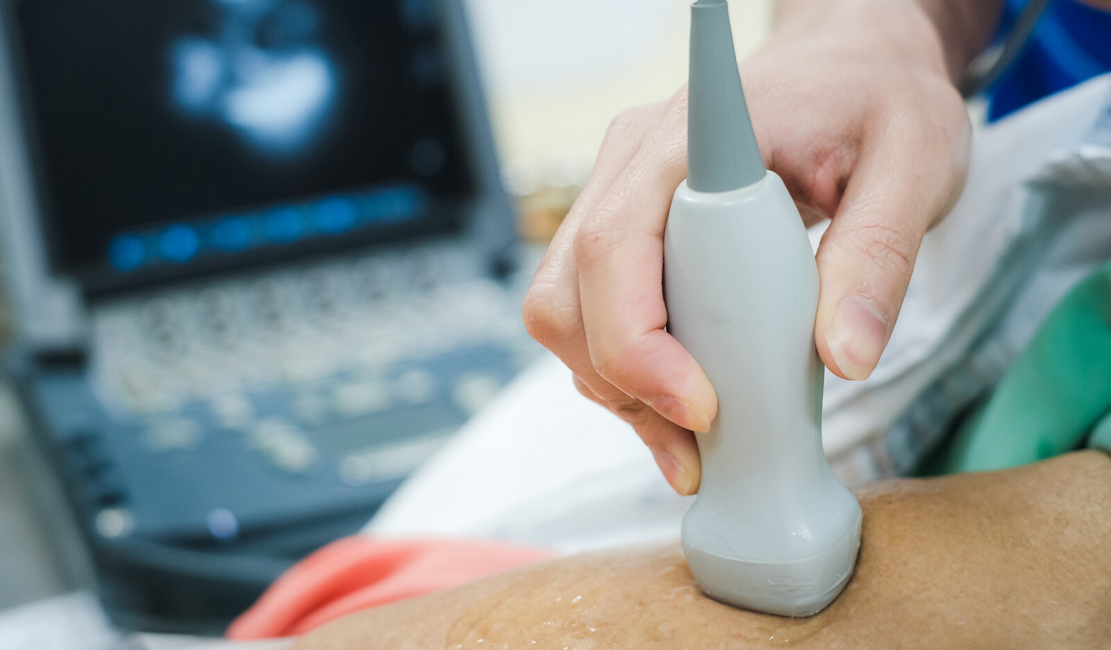
Point of care ultrasound assessment and reporting: Patients randomly allocated to the POCUS group underwent bedside focused sonographic assessment of the heart, lungs and inferior vena cava within 1 h from randomization and within 24 h from ward admission and after the primary team’s first patient assessment. The exam was done according to the local point-of care ultrasonography protocol, which included:
- Cardiac assessment using standard transthoracic echocardiography and inferior vena cava views evaluating ventricular and valvular function and abnormalities, volume status and presence of pericardial fluid
- Lung and pleural space assessment searching for B-lines, pleural effusion or signs of atelectasis or lung consolidation
- Screening for peritoneal fluid
The POCUS operators were not part of the internal ward medical team that took care of the patient and were blinded to the patients’ management plan. During the study year, there were scarce POCUS capabilities among internal ward physicians in this medical center, enabling recruitment of patients naïve to early POCUS assessment.
On completion of the POCUS exam, a full report was completed and handed to the primary physician in the admitting internal ward. The same report was added to the patients’ notes in the electronic medical record, visible to all teams caring for the patient. No diagnostic or treatment recommendations were given by the research team. Primary team exposure to the POCUS report was the only variable differing between the two study arms.
Outcomes: The primary outcomes were time needed to achieve a correct primary diagnosis and time to appropriate treatment given to patients in both study arms. For this purpose, two independent, senior internal medicine experts, with >3 y of cardiac and lung POCUS qualification and experience, separately reviewed patients’ medical records (including imaging modalities, consults and labs). The reviewers did not take part in research design or data collection and did not participate in the POCUS evaluation of the patients in the study.
RESULTS
64 patients met inclusion criteria; 60 were included in the final analysis, and 30 patients were enrolled in each study arm (4 patients declined to participate).
There were no significant differences between the two study groups with respect to age, sex, medical history and initial primary diagnosis. The mean age was 67.8 § 15.3 y in both groups.
POCUS findings and effect on diagnosis and management
Mean time from hospital admission to POCUS scan was 12 h, and all scans were completed within 24 h of hospital admission.
Overall, 22 of 30 patients had abnormal POCUS findings; POCUS evaluation revealed left ventricle dysfunction among 16%, valvulopathy among 30% and lung edema among 13% of patients. Previously unknown clinically relevant findings were detected by POCUS exam among 79% of cases and POCUS findings that led to an additional diagnosis or alteration of previous diagnosis were obtained among 13.8% and 27.6% of patients, respectively.
POCUS effect on clinical outcomes
Equal rates of correct diagnosis were achieved by the primary team in both study arms (80.0% vs. 73.3%, p = 0.543, intervention group vs. control group, respectively). Survival analysis revealed that time needed to achieve a correct diagnosis (as determined by the reviewers) was shorter in the POCUS group than the control group (median time =24 h [95% confidence interval: 18-30 h] vs. 48 h [20-76 h]), but this did not reach statistical significance.
A higher percentage of patients received appropriate treatment in the POCUS group compared with the control group Time to appropriate treatment was about 20 h shorter among patients who underwent early POCUS scan as opposed to patients in the control group (median time of 5 h [95% confidence interval: 0.5-9 h] vs. 24 h [19-29 h].
CONCLUSIONS
The authors concluded that incorporation of the POCUS exam into the early diagnostic routine workup of patients admitted to the medical ward with chest pain or dyspnea reduces time to appropriate therapy.
 English
 Español

