STATE OF THE ART REVIEW | Usefulness of Stress Echocardiography in the Management of Patients Treated with Anticancer Drugs
Source: JASE 2021 34 (2); 107-16
INTRODUCTION
Advances in antineoplastic therapeutic protocols led to improved survival of patients with cancer. This resulted in increased burden of cardiovascular (CV) complications related to cancer treatment. Consequently, a new branch of cardiology has been created, ‘‘cardio-oncology,’’ with the aims of preventing CV complications related to antineoplastic treatment, achieving early diagnosis and treatment of any complications, and allowing completion of the expected antineoplastic treatment.
The development of left ventricular (LV) dysfunction is one of the most serious complications along with QT interval prolongation and arrhythmias, coronary artery disease (CAD), peripheral artery disease and stroke, valvular heart disease and others.
Stress echocardiography has a pivotal role in achieving a timely diagnosis of CAD and thus assisting in management in this clinical setting. The atherosclerotic process can be accelerated by both chemotherapy and chest irradiation in patients with cancer, even several years after anticancer treatment completion. Therefore, the objective of this review was to delineate the role of stress echocardiography in the assessment of CAD in patients with cancer.
DETECTION OF CARDIOTOXICITY
The risk for developing cardiotoxicity is influenced by several conditions, such as patient related factors (i.e., age, preexisting CV disease, baseline global CV risk) and treatment-related factors (type and dose of the scheduled treatment, exposure to previous anticancer treatments and/or to radiotherapy).
Hence, the goal of a cardiooncologist is to stratify the risk for developing cardiac damage from anticancer treatment and a personalized diagnostic and therapeutic workup according to the patient’s risk profile. Assessment of LV function can be performed using several techniques such as echocardiography, cardiac magnetic resonance, and radionuclide multigated acquisition scanning. Echocardiography is the first-line technique because of its wide availability, repeatability, high cost-effectiveness, and lack of exposure to radiation.
Anticancer therapy–related cardiac dysfunction is defined as a decrease in the LV ejection fraction (LVEF) of >10% points to a value less than the lower limit of normal in repeated studies. This value should be confirmed in a measurement repeated after 2 to 3 weeks. The assessment of regional wall motion abnormalities is also valuable. Subtle signs of global and regional dysfunction can be detected using strain imaging analysis. Speckle-tracking echocardiography is a noninvasive ultrasonic technique that allows an objective and quantitative evaluation of both global and regional myocardial function. Particularly global longitudinal strain (GLS), evaluated as an average of the systolic strain values in all myocardial segments, can be used in routine clinical practice and was shown to be a powerful prognostic marker. It has been demonstrated that a reduction in GLS precedes the reduction in LVEF in patients treated with anticancer drugs.
STRESS ECHOCARDIOGRAPHY IN CARDIO-ONCOLOGY
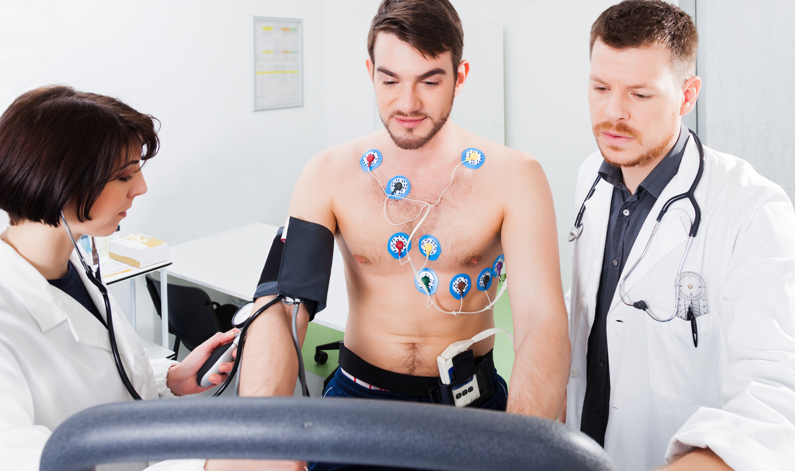
Stress echocardiography provides a dynamic assessment of global and regional LV function under conditions of physiologic (i.e., during exercise) or pharmacologic (i.e., inotropic agents, vasodilators) stress, and it has the advantage of being able to identify any structural or functional anomalies not evident at rest.
In addition, a growing body of evidence has demonstrated many other applications for stress echocardiography, such as in valve disease, systolic and diastolic heart failure, and congenital heart disease. Lastly, another potential application is in cardio-oncology. Anticancer drugs can cause a reduction in LV reserve due to a direct effect on the myocardium or can favor myocardial ischemia inducing accelerated atherosclerosis and endothelial dysfunction or vasospasm. In patients with suspected or diagnosed heart failure, it is useful to evaluate LV contractile reserve (LVCR) by evaluating the increase in LVEF at peak stress compared with baseline and LV elastance (the ratio between blood pressure and end-systolic volume at peak stress and rest).
LVCR is a recognized parameter that predicts survival in patients with CAD. dobutamine stress echocardiography (DSE) can provide information on LVCR in patients with cancer. it appears to be a useful tool for an early identification of oncology patients who are more prone to develop late cardiac dysfunction after anthracycline treatment, when reduction in resting LVEF or diastolic dysfunction is still not evident.
Another interesting tool in patients treated with anticancer drugs could be the noninvasive evaluation of coronary flow reserve (CFR) in the left anterior descending coronary artery (LAD) during dipyridamole stress echocardiography. CFR is the ratio between intra-coronary mean velocity under baseline conditions and after pharmacologic induction of maximum hyperemia. This is a feasible method that, combined with the assessment of wall motion, makes it possible to better stratify patients at high risk for coronary events.
CONCLUSION
The authors point out that as the number of patients surviving cancer continues to increase, cardiac surveillance of these patients is becoming more important. Therefore, it is important that cardiologists and oncologists collaborate to prevent or reduce the occurrence of CV complications of antineoplastic treatment. Therefore, stress echocardiography should be considered for patients undergoing treatment with drugs that induce vascular damage or myocardial damage.
 English
 Español

