BRIEF RESEARCH COMMUNICATIONS • Improved Thrombus Assessment by Transesophageal Echocardiography: The DOLOP (Detection of Left Atrial Appendage Thrombosis Utilizing Optison) Study
Source: JASE 2021:34(8);916-917
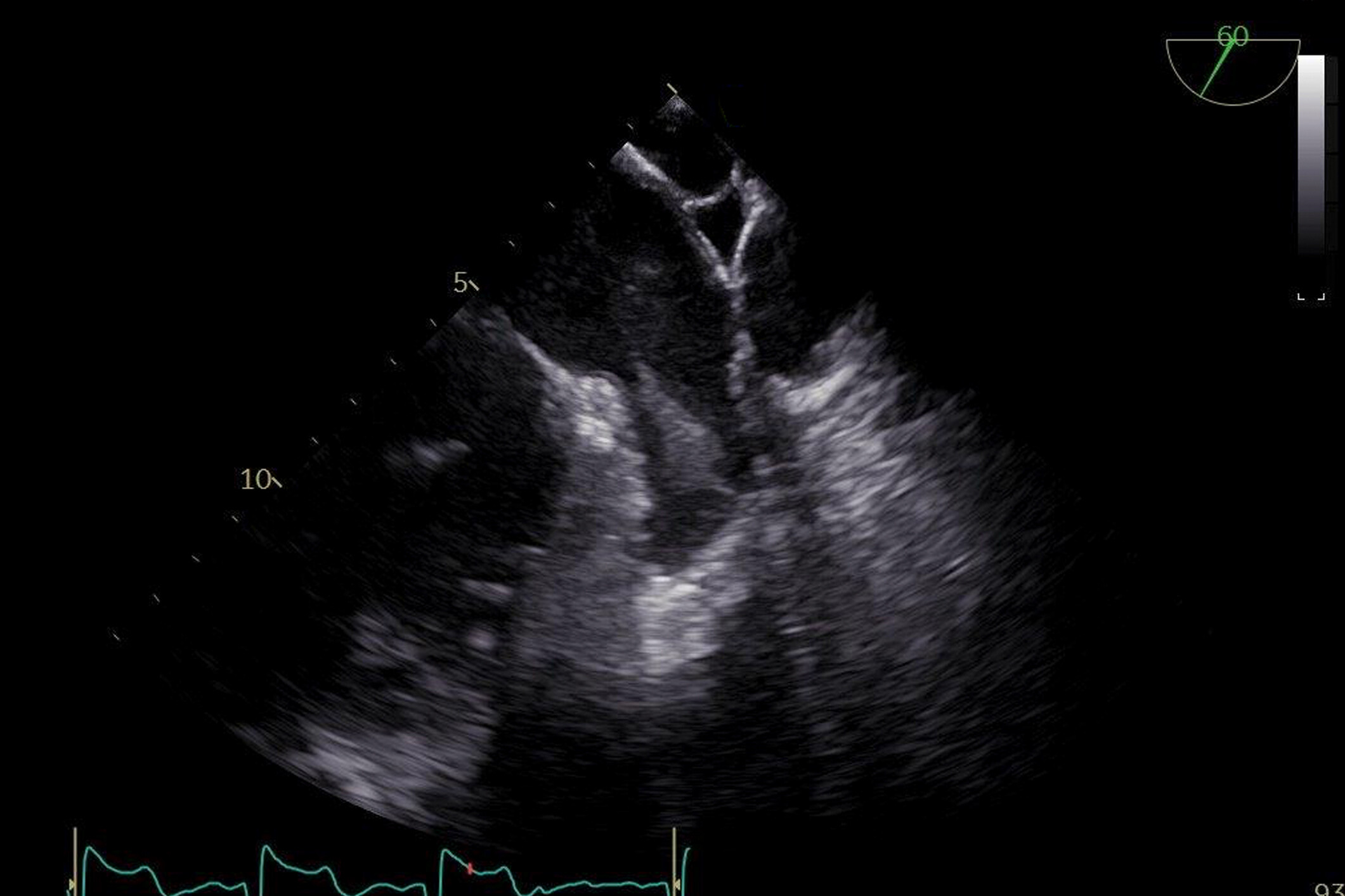
Transesophageal echocardiography (TEE) is an essential clinical tool to assess for thrombus in the left atrial appendage (LAA) prior to cardioversion or ablation in the setting of atrial fibrillation (AF) or flutter. Noncontrast images can be limited by artifact and reverberations, and therefore use of ultrasound-enhancing agents (UEAs) can improve diagnostic accuracy.
The only approved use of UEAs by the Food and Drug Administration is left ventricular opacification. Therefore, use of UEAs in LAA anatomic assessment during TEE is off label. Optison microspheres consist of a serum albumin shell containing perflutren and are less stable when compared with other UEAs. They may offer benefit compared to other UEAs due to shorter persistence which allows for titration.
The authors hypothesized that Optison would improve diagnostic accuracy and certainty in detecting LAA thrombus versus standard TEE evaluation.
The authors prospectively evaluated 428 patients with AF or atrial flutter who presented for TEE to assess for LAA thrombus. Optison was administered if there was increased spontaneous echo contrast (SEC) or uncertainty of LAA thrombus (100 of 428 cases).
Two board-certified echocardiographers independently reviewed paired deidentified pre- and post-UEA image sets. Confidence in presence or absence of thrombus was rated on a 5-point scale for pre- and postcontrast images and as an overall assessment (no confidence, 25%, 50%, 75%, or 100% confidence in presence/absence of thrombus).
Clinical suspicion or poor visualization existed for 100 patients, leading to the use of UEA. Pre-UEA, a thrombus was suspected in 46/100 and 22/100 cases, respectively. Post-UEA thrombus was diagnosed in 14/100 and 10/100 cases, respectively. Four cardioversions, two AF ablations, and one LAA occlusion device implantation were subsequently cancelled.
Agreement between echo readers significantly increased from 72% precontrast (k = 0.414, consistent with moderate agreement) to 96% postcontrast (k = 0.811, almost perfect agreement).
The author concluded that the administration of UEA can lead to significant reduction in potentially inaccurate overreporting of LAA thrombi and improved reader agreement. They believe that the clinical implications of this improved diagnostic accuracy are enormous since the false-positive reporting of thrombus lead to cancellations of procedures (e.g., cardioversions and ablations), repeat imaging, and potentially unnecessary prolonged anticoagulation.
 English
 Español

