STRAIN ECHOCARDIOGRAPHY IN AORTIC STENOSIS • Multichamber Strain Characterization Is a Robust Prognosticator for Both Bicuspid and Tricuspid Aortic Stenosis
Source: J Am Soc Echocardiogr 2022;35:956-65
INTRODUCTION
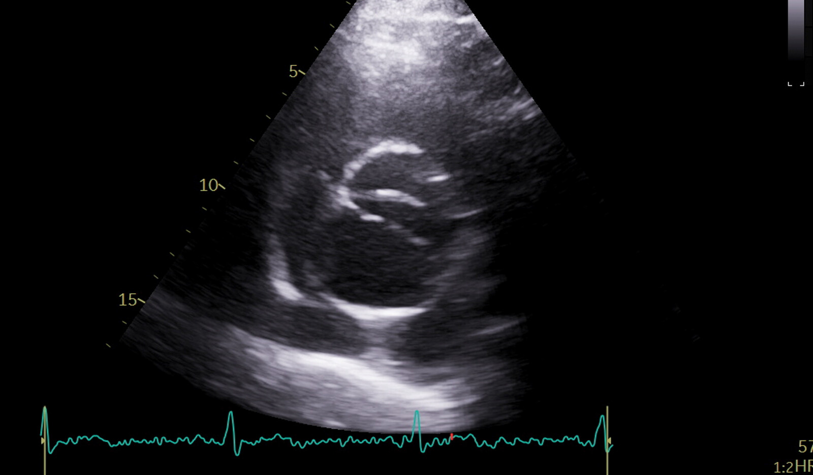
Bicuspid aortic valve (BAV) is the most common congenital cardiac abnormality. It is detected in up to 50% of patients requiring aortic valve replacement (AVR) because of significant aortic stenosis (AS). Patients with BAV-AS are two decades younger, have fewer comorbidities, and exhibit significantly better survival than those with tricuspid aortic valve AS (TAV-AS).
The occurrence of cardiac damage (CD) is progressive in both patients with BAV-AS and those with TAV-AS, with the latter group developing CD at a significantly higher rate than those with BAV. In addition, BAV morphology exhibit an age and comorbidity-independent ‘‘protective’’ effect on CD development in AS. Whether BAV confers ‘‘protection’’ against AS-related CD, or whether subclinical cardiac abnormalities in BAV-AS are overlooked by conventional echocardiographic parameters, is not clear.
The authors hypothesized that patients with more than moderate AS (i.e., moderate to severe AS and greater) may develop subclinical ‘‘downstream’’ CD captured by multichamber strain (MCS) analysis.
Therefore, the aim of this study was to:
- Characterize and compare the extent of impaired longitudinal strain of three chambers (left ventricle, left atrium, and right ventricle) in BAV-AS versus TAV-AS and its relation to CD staging
- Evaluate the prognostic value of MCS in BAV-AS and TAV-AS and whether it is independent of CD staging
METHODS
Study Population
Patients were from an AS cohort designed to compare disease progression and outcomes between BAV-AS (consecutive patients between 2000 and 2013, n = 330) and TAV-AS (1:2 matched to the BAVAS group by year of baseline transthoracic echocardiographic examination, n = 581) with more than mild AS at baseline.
All the patients were retrospectively identified from the echocardiography database. Inclusion criteria included:
- Patients with greater than moderate AS on the last echocardiogram
- Aortic valve [AV] mean gradient [MG] > 30 mm Hg or AV area [AVA] <0 cm2 if stroke volume index < 35 mL/m2)
209 patients with BAV-AS and 384 patients with TAV-AS were left for analysis (time period of echocardiographic examinations, 2004–2018; 90% after 2009). Images eligible for strain analysis were from 100% of patients with BAV-AS and 96% of those with TAV-AS, resulting in 578 total patients for the final analysis.
Clinical and Echocardiographic Variables
Demographic information was ascertained at the time of last transthoracic echocardiographic evaluation. Medical history was retrieved from the electronic medical record. Two-dimensional and Doppler echocardiographic images were clinically acquired by credentialed sonographers and reviewed by level 3–trained, board-certified cardiologists, according to the current American Society of Echocardiography and the European Association of Cardiovascular Imaging guidelines. AVA was calculated using the continuity equation.
Two-Dimensional Semiautomated Strain Analysis
Offline semiautomated strain analyses were performed using the vendor-independent speckle-tracking strain software (2D CPA; TomTec Imaging System). The software traced the LV endocardial border automatically in similar cardiac cycles in apical two chamber, four-chamber, and long-axis views, which was then finetuned manually at end-diastole and end-systole.
LV-GLS was calculated as the average of global strain of three apical views. For LA and RV strain analysis, the software automatically identified the endocardium of the left atrium and right ventricle after manually selecting three points of each chamber.
Outcomes
The primary outcome was the incidence of all-cause mortality.
RESULTS
Patient Characteristics
Most patients had severe AS (94%) and LV ejection fraction > 50% (90%).
Compared with those with TAV, patients with BAV were more than a decade younger, more were men, and they had less cardiovascular risk burden and fewer comorbidities.
The severity of valvular obstruction (percentage of severe AS, AV peak velocity, MG) was similar between BAV and TAV.
After adjustment for age, patients with TAV had more frequent LV hypertrophy, diastolic dysfunction, and higher RV systolic pressure, possibly related to their higher comorbidity burden.
MCS Parameters in BAV-AS versus TAV-AS
Compared with those with TAV-AS, patients with BAV-AS had better global longitudinal strain (LV-GLS), right ventricular free wall longitudinal strain (RVFW-LS), and left atrial reservoir strain (LASr), which were clinically and statistically significant and age independent.
LV-GLS showed moderate to strong correlations with LASr (r = 0.60 [BAV-AS] vs 0.74 [TAV-AS]) and with RVFW-LS (r = 0.63 [BAV-AS] vs 0.71 [TAV-AS]).
LV-GLS was an important single parameter, determining 30% to 50% of variation in measured LASr and RVFW-LS (linear regression adjusted R 2 = 0.3-0.5), suggesting that LV systolic function is a partial determinant of LA reservoir function and RV systolic function in both TAV-AS and BAV-AS.
Outcomes
Five-year survival in the BAV and TAV groups was 86.8 6 2.4% and 49.6 6 2.7%, respectively (log-rank P < .001).
385 patients (85% of those with BAV vs 56% of those with TAV) had AVR. Time from echocardiographic evaluation to AVR was similar in both groups. AVR showed similarly protective effect in the BAV-AS and TAV-AS groups. By landmark analysis, in TAV-AS, AVR showed a favorable effect on survival independent of the number of chambers with impaired strain.
In BAV-AS, AVR showed a protective effect in patients with more than a single chamber with impaired strain. However, the survival improvement afforded by AVR for both groups was less prominent in patients with more chambers with impaired strain, suggesting that more extensively impaired strain might offset the survival benefit of AVR.
Associations of Strain Parameters and MCS with All Cause Death
Three strain parameters, separately, showed significant associations with death either as a continuous or as a dichotomized variable (impaired vs preserved) in both BAV-AS and TAV-AS. This associations remained intact after additional adjustment for CD staging in both groups.
After comprehensive adjustment including CD staging, in BAV-AS, triple-chamber impairment remained significantly associated with worse survival.
In TAV-AS, single-, paired- and triple-chamber impaired strain showed a stepwise increased risk for death compared with preserved strain of all three chambers.
CONCLUSION
The author concluded that MCS characterization is simple, reproducible, and independent of CD staging for prognostication of both patients with BAV and those with TAV with more than moderate AS (>90% severe). They added that echocardiographic strain-centric approach has the potential utility of improving risk stratification and aiding clinical decision-making in AS management, in both patients with BAV AS and those with TAV AS.
 English
 Español

