Image of TTE chest x ray    Online PoCUS Training

Tag Archives: lung ultrasound

Home Posts tagged "lung ultrasound"
Image of TTE chest x ray    Online PoCUS Training

ORIGINAL ARTICLE • Retrospective Analysis of the Diagnostic Accuracy of Lung Ultrasound for Pulmonary Embolism in Patients with and without Pleuritic Chest Pain

Pleuritic chest pain – sharp chest pain exacerbated by breathing or coughing – is a common presenting symptom in the emergency department (ED) and requires a careful differential diagnosis between benign conditions such as musculoskeletal pain, and more serious diseases like pulmonary embolism (...
Read More
Image of TTE chest x ray    Online PoCUS Training

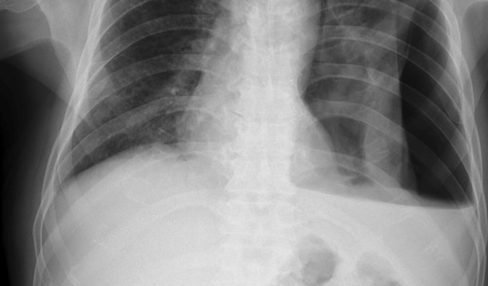
CASE STUDY • ‘Hydro-point’ – The Forgotten and Unspoken Entity in Hydropneumothorax

Lung point-of-care ultrasound (POCUS) showed hydro-point, dynamic air–fluid interface (DAFI) sign and a characteristic triple sign, namely barcode sign/stratosphere sign, hydro-point and sinusoidal pattern in right lung. These features are seen in effusive pneumothorax......
Read More
Image of TTE chest x ray    Online PoCUS Training

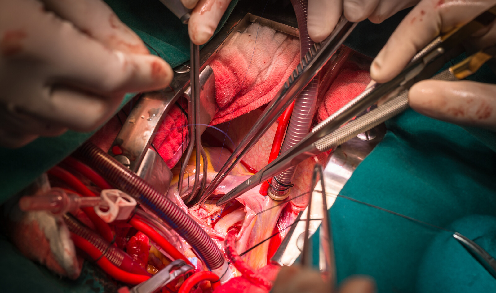
Ventricular Septal Defect Area by Three-Dimensional Assessment of Shunt Severity in Children

Ventricular septal defect (VSD) size is a determinant of shunt severity in children. When compared to three-dimensional (3D) echocardiography (3DE) and surgical measures, Two-dimensional (2D) echocardiography (2DE) underestimates VSD size ......
Read More
Image of TTE chest x ray    Online PoCUS Training

ORIGINAL ARTICLE • Lung Ultrasound Training and Evaluation for Proficiency Among Physicians in a Low-Resource Setting

Lung ultrasound (LUS) is an effective tool to evaluate patients with dyspnea in the emergency department (ED). While a systematic review of LUS training recommended a three-step approach of teaching theoretical knowledge ......
Read More
Image of TTE chest x ray    Online PoCUS Training

RESEARCH • Lung Ultrasound in a Tertiary Intensive Care Unit Population: A Diagnostic Accuracy Study

Etiology of respiratory failure in ICU patients include cardiogenic pulmonary edema (CPE), acute respiratory distress syndrome (ARDS), atelectasis, and pneumonia . Chest X-ray and thoracic computed tomography (CT) are still the most common diagnostic modalities of ICU patients with respiratory failu...
Read More
Image of TTE chest x ray    Online PoCUS Training

ORIGINAL ARTICLE • Results from a Spanish National Survey on the Application of Ultrasound in Pulmonology Services

While ultrasound use initially started for guidance during pleural procedures, over time it has proven useful in the assessment of the lung parenchyma, diaphragm, and chest wall. Therefore, it has become a basic first-line tool to establish a differential diagnosis in patients with dyspnea......
Read More
Image of TTE chest x ray    Online PoCUS Training

ECHOCARDIOGRAPHY IN COVID-19 • The Predictive Role of Combined Cardiac and Lung Ultrasound in Coronavirus Disease 2019

Despite the recognition of the prognostic significance and clinical implications of the cardiac complications of coronavirus disease 2019 (COVID-19), both the European Association of Cardiovascular Imaging and the American Society of Echocardiography recommend limiting the echocardiographic assessme...
Read More
Image of TTE chest x ray    Online PoCUS Training

Research Letter: Lung Ultrasound Can Predict Response to the Prone Position in Awake Non-Intubated Patients with COVID-19 Associated Acute Respiratory Distress Syndrome.

Prone positioning (PP) is often used in acute respiratory distress syndrome (ARDS). While studies have demonstrated positive effects on oxygenation in awake and non-intubated patients, it isn't effective in every case......
Read More
Image of TTE chest x ray    Online PoCUS Training

Review: Role of Point-of-Care Ultrasound During the COVID-19 Pandemic: Our Recommendations in the Management of Dialytic Patients

LUS is well validated in evaluation of several respiratory conditions. LUS is an image method that classically allows the evaluation of the pulmonary periphery, which is helpful in assessing COVID-19.......
Read More