All posts by emedv2b_zvusge

Página de inicio Articles posted by emedv2b_zvusge (Page 3)
Image of skelatal muscle recruitable perfusion LVAD Left Ventricular Assist Device Implantation contrast ultrasound    Online PoCUS Training

Right Ventricular Postsystolic Strain Curve Morphology before and after Vasodilator Treatment in Idiopathic Pulmonary Arterial Hypertension

The authors discussed the current guidelines for diagnostic workup of pulmonary hypertension, which include right heart catheterization to establish diagnosis, a transthoracic echocardiography to rule out left heart disease, a ventilation/perfusion lung scan to exclude chronic thromboembolic pulmona...
Read More
Image of skelatal muscle recruitable perfusion LVAD Left Ventricular Assist Device Implantation contrast ultrasound    Online PoCUS Training

ECHOCARDIOGRAPHY AND PULMONARY HYPERTENSION • Echocardiographic Biventricular Coupling Index to Predict Precapillary Pulmonary Hypertension

Right heart catheterization (RHC) remains the gold standard in the diagnostic workup. However, transthoracic echocardiography is currently part of the screening process and is increasingly used in the longitudinal follow-up of patients with PH. Estimated systolic pulmonary artery pressure (sPAP) is ...
Read More
Image of skelatal muscle recruitable perfusion LVAD Left Ventricular Assist Device Implantation contrast ultrasound    Online PoCUS Training

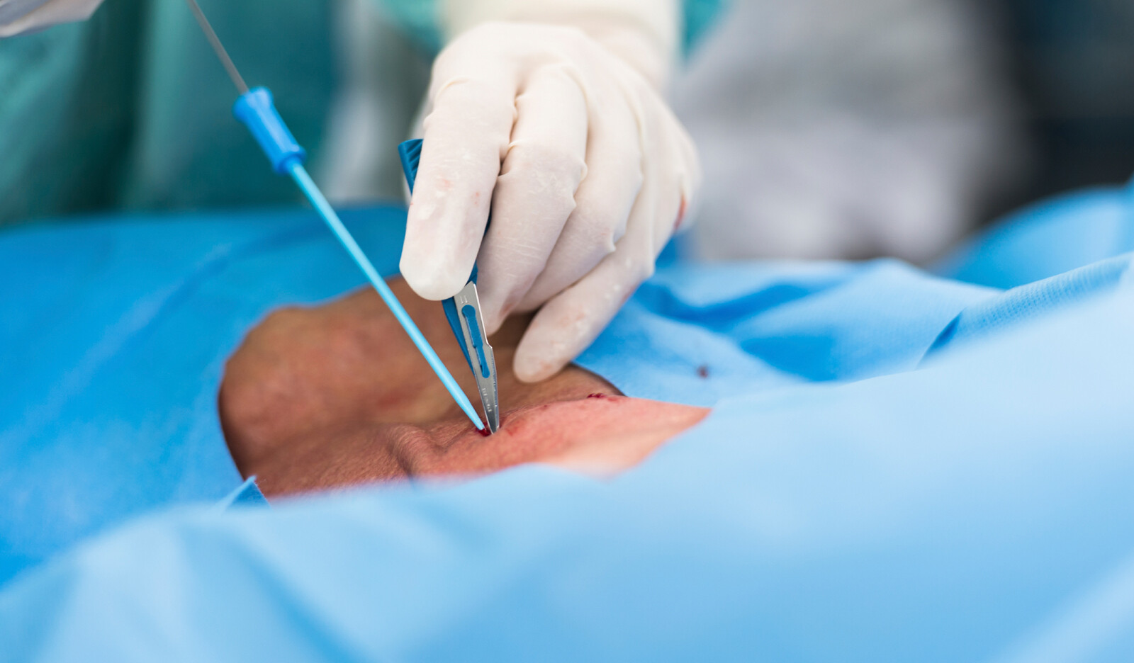
ORIGINAL ARTICLE • Prehospital Portable Ultrasound for Safe and Accurate Prehospital Needle Thoracostomy: A Pilot Educational Study

Since prehospital POCUS is becoming more widely adopted for its diagnostic abilities to aid in decision-making in traumatically injured patients, the authors designed this pilot educational study intervention to train ALS prehospital personnel to use a handheld POCUS to aid in the detection of pneum...
Read More
Image of skelatal muscle recruitable perfusion LVAD Left Ventricular Assist Device Implantation contrast ultrasound    Online PoCUS Training

ORIGINAL ARTICLE • Relationship of the Internal Jugular Vein to the Common Carotid Artery: Implications for Ultrasound-Guided Vascular Access

The internal jugular vein (IJV) is a commonly used cannulation site for central venous access as it is easily accessible due to its superficial anatomical position. The traditional landmark-based technique for IJV cannulation depends on it having a lateral position relative to the common carotid art...
Read More
Image of skelatal muscle recruitable perfusion LVAD Left Ventricular Assist Device Implantation contrast ultrasound    Online PoCUS Training

PERICARDIAL FAT AND CARDIAC FUNCTION • Association of Pericardial Fat with Cardiac Structure, Function, and Mechanics: The Multi-Ethnic Study of Atherosclerosis

Among 6,814 participants initially recruited in MESA, the authors included the 3,032 participants who underwent echocardiography at exam 6. At baseline, this cohort was 53% female, with mean age 57 years, and 40% White, 25% Black or African American, 22% Hispanic, and 13% Asian......
Read More
Image of skelatal muscle recruitable perfusion LVAD Left Ventricular Assist Device Implantation contrast ultrasound    Online PoCUS Training

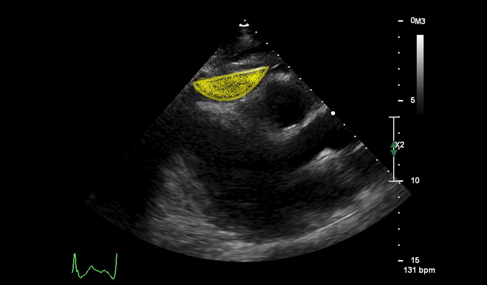
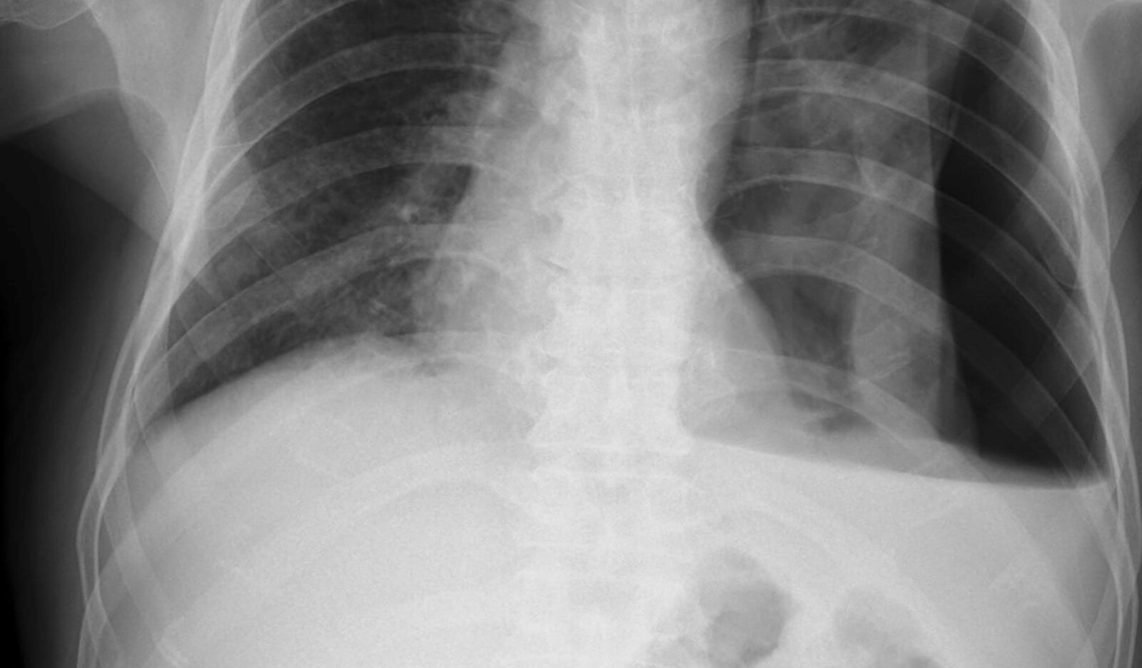
CASE STUDY • ‘Hydro-point’ – The Forgotten and Unspoken Entity in Hydropneumothorax

Lung point-of-care ultrasound (POCUS) showed hydro-point, dynamic air–fluid interface (DAFI) sign and a characteristic triple sign, namely barcode sign/stratosphere sign, hydro-point and sinusoidal pattern in right lung. These features are seen in effusive pneumothorax......
Read More
Image of skelatal muscle recruitable perfusion LVAD Left Ventricular Assist Device Implantation contrast ultrasound    Online PoCUS Training

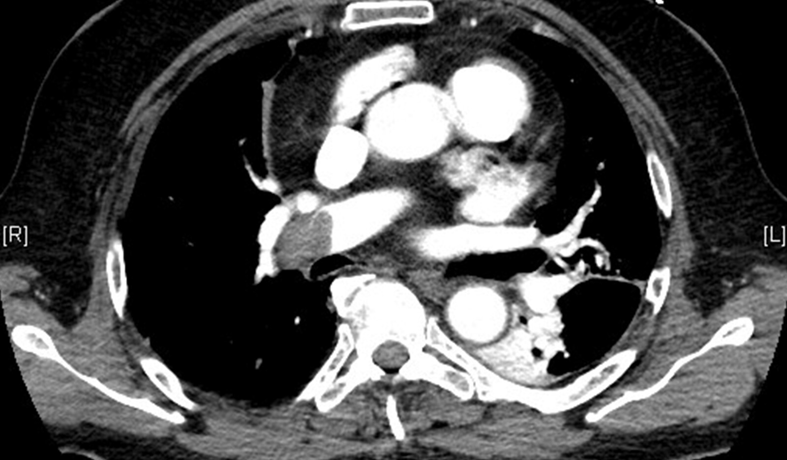
RESEARCH • Intermediate-Risk Pulmonary Embolism: Echocardiography Predictors of Clinical Deterioration

Acute pulmonary embolism (PE) can lead to clinical deterioration due to its effect on the right ventricle (RV). Since there are no consistent definitions or assessments of abnormal RV (abnlRV) there is limited data on association between abnlRV and clinical outcomes and prognostic performance......
Read More
Image of skelatal muscle recruitable perfusion LVAD Left Ventricular Assist Device Implantation contrast ultrasound    Online PoCUS Training

PERICARDIAL FAT AND CARDIAC FUNCTION • Association of Pericardial Fat with Cardiac Structure, Function, and Mechanics: The Multi-Ethnic Study of Atherosclerosis

The authors utilized data from the Multi-Ethnic Study of Atherosclerosis (MESA) to examine the association between pericardial adipose tissue volume by computed tomography (CT) with cardiac structure and function by echocardiography......
Read More
Image of skelatal muscle recruitable perfusion LVAD Left Ventricular Assist Device Implantation contrast ultrasound    Online PoCUS Training

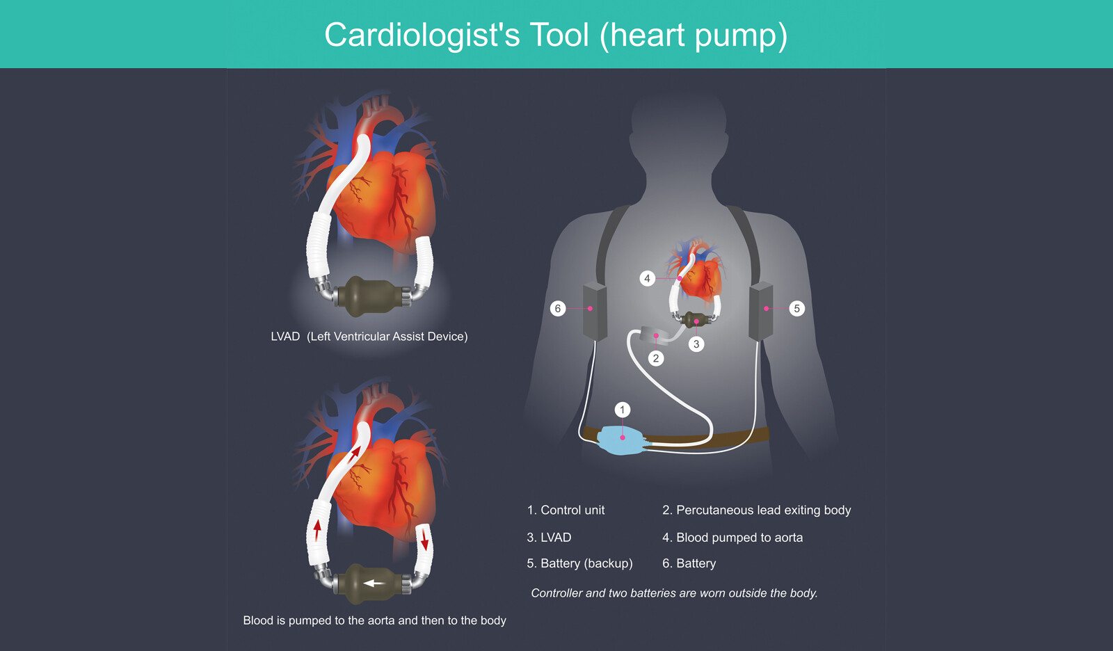
KELETAL MUSCLE PERFUSION DURING MECHANICAL CIRCULATORY SUPPORT • Contrast Ultrasound Assessment of Skeletal Muscle Recruitable Perfusion after Permanent Left Ventricular Assist Device Implantation: Implications for Functional Recovery

While diagnosis can be made with computed tomography, magnetic resonance imaging, cardiac catheterization, and echocardiography, only echocardiography which has the advantage of low cost and good safety profile, is convenient for use as a screening tool......
Read More